
Distal radial fracture
Updates to Article Attributes
This is a basic article for medical students and other non-radiologists
Distal radial fractures usually are a heterogeneous group of fractures that occur at the distal radius and are the dominant fracture type at the wrist.
They usually occur when significant force is applied to the distal radial metaphysis. Fracture occurs when the force applied is greater than the force that can be absorbed by the bone.
They are best described in terms of their fracture type, location, displacement and joint involvement. Traditionally, eponymous names were given to the common fracture types of the distal radius:
- Colles fracture: transverse extra-articular fracture with dorsal angulation
- Smith fracture: transverse fracture with palmar angulation
Background
Relevant anatomy
The forearm has a patient fallspaired radius and ulna which are both wedge-shaped. The radius is narrow at the radial head (elbow) and wide at its articulation with the carpus (at the wrist). The ulna has the opposite configuration.
This means that any longitudinal force placed through the hand and wrist is preferentially absorbed by the radius.
Relevant physiology
By adolescence the bones of normal people are strong and the physes are fused. To fracture the bone of a young person requires a significant amount of energy.
As people age, their bone density (and therefore bone strength) decreases. Reduction of bone mineral density is greater in women, particularly if they are on hormone replacement therapy. Other drugs such as steroids can also have a negative effect on bone density.
Demographics
Distal radial fractures can be seen in any group of patients and there is a bimodal age and sex distribution. Younger patients tending to be male and older patients tending to be female.
Aetiology
Trauma is almost always the cause of distal radial fractures and is often the result of a fall onto an outstretched hand (FOOSH).
Content coming
In the elderly, the bones tend to have a much lower bone density and are consequently much weaker. Fracture of the distal radius can occur with injuries that exert much less force, e.g. falling from standing height.
Pathophysiology
Force applied longitudinally or obliquely to the hand and wrist is absorbed by the distal radius because it is the load-bearing bone in the forearm. If this force is greater than the strength of the bone, a fracture occurs.
When most people fall, they don't axially load the forearm, but apply an oblique force longitudinally and dorsally. If a fracture does occur, there is usually associated dorsal angulation.
Classification
There are many radiological classification systems - probably a sign that no one single classification system works. It is more important to recognise what makes the fracture worse:
- oblique, spiral or comminuted configuration
- greater degree of angulation
- intra-articular involvement
- additional fractures
Clinical features
Presentation
The majority of patients with a distal radial fracture present following a fall onto an outstretched hand. They are in pain and have a reduced range of motion. There may be an associated deformity and in severe cases, distal neurovascular compromise.
Diagnosis
Diagnosis usually only requires a standard wrist x-ray (AP and lateral film). In some complex cases, additional cross-sectional imaging (CT scan) is required to accurately assess the fracture. This is especially true when there is a multi-part fracture with joint involvement (uncommon).
Treatment and prognosis
Treatment is dependant on the type of fracture (as determined by the x-ray). The vast majority of distal radial fractures are relatively uncomplicated and do not require a trip to theatre and can be managed as an out-patient with review in fracture clinic.
Fractures with significant displacement require manipulation (in A&E under sedation or theatre under anaesthetic). A small number will require internal fixation following manipulation.
A small proportion of patients treated conservatively need to be followed up. If a fracture is stable and treated in cast it must be reviewed regularly because of the risk of displacement. This is particularly true if the cast becomes loose once the wrist swelling subsides. Late displacement warrants surgical consideration.
Radiographic features
X-ray features
Most distal radial fractures in adult patients are transverse metaphyseal fractures. They are often extra-articular, but some may extend into the joint and when they do, it is important to recognise. The degree of displacement (usually dorsal) is important because it will be a determining factor for treatment (whether to reduce or not before immobilisation).
When describing the fracture, think about:
- what type of fracture is it?
- is there displacement?
- is there joint involvement?
- is there an accompanying ulnar styloid fracture?
-<h6>This is a basic article for medical students and non-radiologists</h6><p><strong>Distal radial fractures</strong> usually occur when a patient falls onto an outstretched hand (FOOSH). </p><p><em>Content coming</em></p>- +<h6>This is a basic article for medical students and other non-radiologists</h6><p><strong>Distal radial fractures</strong> are a heterogeneous group of fractures that occur at the distal radius and are the dominant fracture type at the wrist. </p><p>They usually occur when significant force is applied to the distal radial metaphysis. Fracture occurs when the force applied is greater than the force that can be absorbed by the bone.</p><p>They are best described in terms of their fracture type, location, displacement and joint involvement. Traditionally, eponymous names were given to the common fracture types of the distal radius:</p><ul>
- +<li>
- +<a href="/articles/colles-fracture">Colles fracture</a>: transverse extra-articular fracture with dorsal angulation</li>
- +<li>
- +<a href="/articles/smith-fracture">Smith fracture</a>: transverse fracture with palmar angulation</li>
- +</ul><h4>Background</h4><h5>Relevant anatomy</h5><p>The forearm has a paired radius and ulna which are both wedge-shaped. The radius is narrow at the radial head (elbow) and wide at its articulation with the carpus (at the wrist). The ulna has the opposite configuration.</p><p>This means that any longitudinal force placed through the hand and wrist is preferentially absorbed by the radius.</p><h5>Relevant physiology</h5><p>By adolescence the bones of normal people are strong and the physes are fused. To fracture the bone of a young person requires a significant amount of energy.</p><p>As people age, their bone density (and therefore bone strength) decreases. Reduction of bone mineral density is greater in women, particularly if they are on hormone replacement therapy. Other drugs such as steroids can also have a negative effect on bone density.</p><h5>Demographics</h5><p>Distal radial fractures can be seen in any group of patients and there is a bimodal age and sex distribution. Younger patients tending to be male and older patients tending to be female.</p><h5>Aetiology</h5><p>Trauma is almost always the cause of distal radial fractures and is often the result of a fall onto an outstretched hand (FOOSH).</p><p>In young adults, the long bones tend to be strong and the force required to break the bone is significant. Thus, distal radial fractures in younger patients require much greater force, e.g. falling from a significant height, severe road traffic accident.</p><p>In the elderly, the bones tend to have a much lower bone density and are consequently much weaker. Fracture of the distal radius can occur with injuries that exert much less force, e.g. falling from standing height.</p><h5>Pathophysiology</h5><p>Force applied longitudinally or obliquely to the hand and wrist is absorbed by the distal radius because it is the load-bearing bone in the forearm. If this force is greater than the strength of the bone, a fracture occurs.</p><p>When most people fall, they don't axially load the forearm, but apply an oblique force longitudinally and dorsally. If a fracture does occur, there is usually associated dorsal angulation.</p><h5>Classification</h5><p>There are many radiological classification systems - probably a sign that no one single classification system works. It is more important to recognise what makes the fracture worse:</p><ul>
- +<li>oblique, spiral or comminuted configuration</li>
- +<li>greater degree of angulation</li>
- +<li>intra-articular involvement</li>
- +<li>additional fractures</li>
- +</ul><h4>Clinical features</h4><h5>Presentation</h5><p>The majority of patients with a distal radial fracture present following a <a href="/articles/investigation-of-fall-onto-an-outstretched-hand-basic">fall onto an outstretched hand</a>. They are in pain and have a reduced range of motion. There may be an associated deformity and in severe cases, distal neurovascular compromise.</p><h5>Diagnosis</h5><p>Diagnosis usually only requires a standard wrist x-ray (AP and lateral film). In some complex cases, additional cross-sectional imaging (CT scan) is required to accurately assess the fracture. This is especially true when there is a multi-part fracture with joint involvement (uncommon).</p><h5>Treatment and prognosis</h5><p>Treatment is dependant on the type of fracture (as determined by the x-ray). The vast majority of distal radial fractures are relatively uncomplicated and do not require a trip to theatre and can be managed as an out-patient with review in fracture clinic.</p><p>Fractures with significant displacement require manipulation (in A&E under sedation or theatre under anaesthetic). A small number will require internal fixation following manipulation.</p><p>A small proportion of patients treated conservatively need to be followed up. If a fracture is stable and treated in cast it must be reviewed regularly because of the risk of displacement. This is particularly true if the cast becomes loose once the wrist swelling subsides. Late displacement warrants surgical consideration.</p><h4>Radiographic features</h4><h5>X-ray features</h5><p>Most distal radial fractures in adult patients are <a href="/articles/transverse-fracture">transverse</a> metaphyseal fractures. They are often extra-articular, but some may extend into the joint and when they do, it is important to recognise. The degree of displacement (usually dorsal) is important because it will be a determining factor for treatment (whether to reduce or not before immobilisation).</p><p>When <a href="/articles/describing-a-fracture-an-approach">describing the fracture</a>, think about:</p><ul>
- +<li>what type of fracture is it?</li>
- +<li>is there displacement?</li>
- +<li>is there joint involvement?</li>
- +<li>is there an accompanying ulnar styloid fracture?</li>
- +</ul>
Systems changed:
- Musculoskeletal
Image ( create )
Image 2 X-ray (Lateral) ( create )
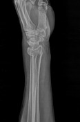
Image 3 X-ray (Lateral) ( create )

Image 4 X-ray (Oblique) ( create )

Image 6 X-ray (Frontal) ( create )


 Unable to process the form. Check for errors and try again.