
Left ventricular ejection fraction (echocardiography)
Updates to Article Attributes
Left ventricular ejection fraction is a surrogate for left ventricular global systolic function, defined as the left ventricular stroke volume divided by the end-diastolic volume.9
Terminology
Point-of-care echocardiography protocols typically use a semi-quantitative approach in defining the ejection fraction, describing the function as hyperdynamic (estimated ejection fraction >70%), normal (55-70%), or depressed (<55%).
If the global function is depressed it is then appropriate to further describe the dysfunction as mild (40-55%), moderate (30-40%), or severe (<30%).
Radiographic features
A global visual assessment of left ventricular systolic function precedes formal quantification of ejection fraction. A gestalt of systolic function may be inferred from the degree of inward endocardial motion, myocardial thickening, longitudinal motion of the mitral annulus, and the overall geometry of the ventricle.
Visual estimates can be accurate as more complex time‐consuming measurements in assessing left ventricular ejection fraction.4,5.
Ultrasound
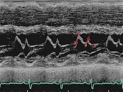
- mitral valve E point septal separation (EPSS, normal < 7 mm)1
- the distance between the anterior mitral valve leaflet and the interventricular septum in early diastole
- an EPSS > 7mm is 100% sensitive for a severely depressed ejection fraction (EF < 30%)7
- a cutoff EPSS > 8 mm was 83.3% sensitive and 50.0% specific for any systolic dysfunction
-
Asas derived from comparison with MRI LVEF, (75.5 x 2.5) x EPSS roughly equates ejection fraction8
- fractional shortening (normal range 25
% – 45-45% )- assessed immediately distal to the tips of the mitral valve leaflets in the parasternal long axis view3
- the ventricular dimensions at end-diastole (LVIDd) and end-systole (LVIDs) are used to calculate a
percentper cent change - FS = (LVIDd - LVIDs) / LVIDd
- fractional shortening x 2 roughly equates ejection fraction
- biplane method of discs (modified Simpson’s rule)
-
Usingusing two orthogonal apical views, the endocardial border is outlined in end-diastole and end-systole - the ventricle is divided along its long axis into a series of disks of equal height, and ventricular volume is calculated as the sum of the volume of the disks
-
- mitral annular plane systolic excursion
- m-mode pick directed at the septal (and/or lateral) mitral annulus
- maximal apical excursion measured in mm
-
Suggestivesuggestive of decreased EF when <11mm;11 mm
- fractional area change (normal range 35-65%)
- the endocardium is traced at end systole and end diastole in the parasternal short axis at the mid-papillary level
- FAC = (LV end-diastolic area - LV end systolic area) / LVEDA
Differential diagnosis
General imaging differential considerations for a reduced ejection fraction include:
- regional wall motion abnormality
- non-perpendicular alignment of the M-mode line in relation to the long axis of the LV
- poor delineation of the endocardial and epicardial borders
- contiguous structures such as the chordae, trabeculations near the posterior wall, and false-tendons may
-
Abnormalabnormal left ventricular geometry- such as aneurysmal
remodelingremodelling
- such as aneurysmal
-
Valvularvalvular pathology- EPSS will not reflect ejection fraction in the presence of significant aortic insufficiency/mitral stenosis
-<p><strong>Left ventricular ejection fraction</strong> is a surrogate for left ventricular global systolic function, defined as the left ventricular stroke volume divided by the end-diastolic volume.<sup>9</sup> </p><h4>Terminology</h4><p>Point-of-care echocardiography protocols typically use a semi-quantitative approach in defining the ejection fraction, describing the function as hyperdynamic (estimated ejection fraction >70%), normal (55-70%), or depressed (<55%).</p><p>If the global function is depressed it is then appropriate to further describe the dysfunction as mild (40-55%), moderate (30-40%), or severe (<30%). </p><h4>Radiographic features</h4><p>A global visual assessment of left ventricular systolic function precedes formal quantification of ejection fraction. A gestalt of systolic function may be inferred from the degree of inward endocardial motion, myocardial thickening, longitudinal motion of the mitral annulus, and the overall geometry of the ventricle.</p><p>Visual estimates can be accurate as more complex time‐consuming measurements in assessing left ventricular ejection fraction.<sup>4,5</sup></p><h5>Ultrasound</h5><ul>-<li>mitral valve E point septal separation (EPSS, normal < 7 mm)<sup>1</sup> <ul>- +<p><strong>Left ventricular ejection fraction</strong> is a surrogate for left ventricular global systolic function, defined as the left ventricular stroke volume divided by the end-diastolic volume.</p><h4>Terminology</h4><p>Point-of-care echocardiography protocols typically use a semi-quantitative approach in defining the ejection fraction, describing the function as hyperdynamic (estimated ejection fraction >70%), normal (55-70%), or depressed (<55%).</p><p>If the global function is depressed it is then appropriate to further describe the dysfunction as mild (40-55%), moderate (30-40%), or severe (<30%). </p><h4>Radiographic features</h4><p>A global visual assessment of left ventricular systolic function precedes formal quantification of ejection fraction. A gestalt of systolic function may be inferred from the degree of inward endocardial motion, myocardial thickening, longitudinal motion of the mitral annulus, and the overall geometry of the ventricle.</p><p>Visual estimates can be accurate as more complex time‐consuming measurements in assessing left ventricular ejection fraction <sup>4,5</sup>.</p><h5>Ultrasound</h5><ul>
- +<li>mitral valve E point septal separation (EPSS, normal < 7 mm) <sup>1</sup> <ul>
-<li>As derived from comparison with MRI LVEF, (75.5 x 2.5) x EPSS roughly equates ejection fraction<sup>8</sup>- +<li>as derived from comparison with MRI LVEF, (75.5 x 2.5) x EPSS roughly equates ejection fraction <sup>8</sup>
-<li>fractional shortening (normal range 25% – 45% )<ul>-<li>assessed immediately distal to the tips of the mitral valve leaflets in the parasternal long axis view<sup>3</sup> </li>-<li>the ventricular dimensions at end-diastole (LVIDd) and end-systole (LVIDs) are used to calculate a percent change</li>- +<li>fractional shortening (normal range 25-45% )<ul>
- +<li>assessed immediately distal to the tips of the mitral valve leaflets in the parasternal long axis view <sup>3</sup> </li>
- +<li>the ventricular dimensions at end-diastole (LVIDd) and end-systole (LVIDs) are used to calculate a per cent change</li>
-<li>Using two orthogonal apical views, the endocardial border is outlined in end-diastole and end-systole</li>- +<li>using two orthogonal apical views, the endocardial border is outlined in end-diastole and end-systole</li>
-<li>Suggestive of decreased EF when <11mm </li>- +<li>suggestive of decreased EF when <11 mm </li>
-<li>Abnormal left ventricular geometry<ul><li>such as aneurysmal remodeling</li></ul>- +<li>abnormal left ventricular geometry<ul><li>such as aneurysmal remodelling</li></ul>
-<li>Valvular pathology<ul><li>EPSS will not reflect ejection fraction in the presence of significant aortic insufficiency/mitral stenosis</li></ul>- +<li>valvular pathology<ul><li>EPSS will not reflect ejection fraction in the presence of significant aortic insufficiency/mitral stenosis</li></ul>
-</ul><p> </p>- +</ul>
Image 1 Ultrasound (Parasternal long axis view) ( update )


 Unable to process the form. Check for errors and try again.