
Organ of Zuckerkandl
Updates to Article Attributes
The Organ of Zuckerkandl comprises of a small mass of chromaffin cells derived from neural crest located along the aorta, beginning cranial to the superior mesenteric artery or renal arteries and extending to the level of the aortic bifurcation or just beyond. The highest concentration is typically seen at the origin of the inferior mesenteric artery.
Physiology
Its physiological role is thought to be of greatest importance duringthe early gestational period as a homeostatic regulator of bloodpressure, secreting catecholamines into the fetal circulation. Theorgan regresses in the end of gestation and following birthto form the aorticosympathetic group of the adult paraganglia.
Radiographic features
The organs of Zuckerkandl are not often visualized radiologically unless they are involved by a pathologic process. Such include
- paragangliomas 1
- neuroblastoma: rare 3
Etymology
It was first described in 1901 by Emil Zuckerkandl, a professor of anatomy at the University of Vienna.
-<p>The <strong>Organ of Zuckerkandl </strong>comprises of a small mass of <a href="/articles/chromaffin-cells" title="Chromaffin cells" style="color: rgb(63, 117, 216); text-decoration: none; ">chromaffin cells</a> derived from neural crest located along the aorta, beginning cranial to the superior mesenteric artery or renal arteries and extending to the level of the aortic bifurcation or just beyond. The highest concentration is typically seen at the origin of the <a href="/articles/inferior-mesenteric-artery" title="Inferior mesenteric artery (IMA) " style="color: rgb(63, 117, 216); text-decoration: none; ">inferior mesenteric artery</a>.</p><h4>Physiology</h4><p>Its physiological role is thought to be of greatest importance during-the early gestational period as a homeostatic regulator of blood-pressure, secreting catecholamines into the fetal circulation. The-organ regresses in the end of gestation and following birth-to form the aorticosympathetic group of the adult <a href="/articles/paraganglia" title="paraganglia">paraganglia</a>.</p><h4>Radiographic features</h4><p>The organs of Zuckerkandl are not often visualized radiologically unless they are involved by a pathologic process. Such include</p><ul>-<li>-<a href="/articles/paraganglioma-1" title="Paraganglioma">paragangliomas</a> <sup>1</sup><ul><li><a href="/articles/pheochromocytoma-2" title="Phaeochromocytoma">phaeochromocytoma</a></li></ul>-</li>-<li>-<a href="/articles/neuroblastoma" title="Neuroblastoma">neuroblastoma</a>: rare <sup>3</sup>-</li>-</ul><h4>Etymology</h4><p>It was first described in 1901 by <strong>Emil Zuckerkandl</strong>, a professor of anatomy at the University of Vienna. </p>- +<p>The <strong>Organ of Zuckerkandl </strong>comprises of a small mass of <a href="/articles/chromaffin-cells">chromaffin cells</a> derived from neural crest located along the <a title="Aorta" href="/articles/aorta">aorta</a>, beginning cranial to the <a title="Superior mesenteric artery" href="/articles/superior-mesenteric-artery">superior mesenteric artery</a> or <a title="Renal arteries" href="/articles/renal-artery">renal arteries</a> and extending to the level of the aortic bifurcation or just beyond. The highest concentration is typically seen at the origin of the <a href="/articles/inferior-mesenteric-artery">inferior mesenteric artery</a>.</p><h4>Physiology</h4><p>Its physiological role is thought to be of greatest importance during the early gestational period as a homeostatic regulator of blood pressure, secreting catecholamines into the fetal circulation. The organ regresses in the end of gestation and following birth to form the aorticosympathetic group of the adult <a href="/articles/paraganglia">paraganglia</a>.</p><h4>Radiographic features</h4><p>The organs of Zuckerkandl are not often visualized radiologically unless they are involved by a pathologic process. Such include</p><ul>
- +<li>
- +<a href="/articles/paraganglioma-1">paragangliomas</a> <sup>1</sup><ul><li><a href="/articles/pheochromocytoma-2">phaeochromocytoma</a></li></ul>
- +</li>
- +<li>
- +<a href="/articles/neuroblastoma">neuroblastoma</a>: rare <sup>3</sup>
- +</li>
- +</ul><h4>Etymology</h4><p>It was first described in 1901 by <strong>Emil Zuckerkandl</strong>, a professor of anatomy at the University of Vienna.</p>
Image 1 MRI (T2 HASTE) ( update )
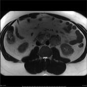
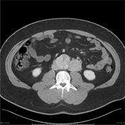
Image 2 CT (C+ portal venous phase) ( update )


 Unable to process the form. Check for errors and try again.