
Peripheral arterial disease
Updates to Article Attributes
Peripheral arterial disease is a common and debilitating condition.
Epidemiology
The age-adjusted prevalence of peripheral arterial disease is approximately 12% 3.
Pathology
Atherosclerosis is the leading cause of occlusive arterial disease of the extremities in patients over 40 years of age with the highest incidence in the sixth and seventh decades of life.
Risk factors
The risk factors for PAD are basically the same as for coronary artery disease:
- diabetes mellitus
- cigarette smoking
- advancing age
- hypercholesterolaemia
- hypertension
- overweight/obesity
Clinical classification
TheRutherford classification
- stage 0: asymptomatic
- stage 1: mild claudication
- stage 2: moderate claudication : the distance that delineates mild, moderate and severe claudication is not specified in the Rutherford classification, but is mentioned in the Fontaine classification as 200 meters
- stage 3: severe claudication
- stage 4: rest pain
- stage 5: ischaemic ulceration not exceeding ulcer of the digits of the foot
- stage 6: severe ischemic ulcers or frank gangrene
TheFontaine classification
- stage I: asymptomatic.
-
stage II: intermittent claudication.
- stage IIa: intermittent claudication after more than 200 meters of pain free walking.
- stage IIb: intermittent claudication after less than 200 meters of walking
- stage III: rest pain.
- stave IV: ischaemic ulcers or gangrene
TASC II classi classification of femoral and popliteal lesions4
-
type A lesions
- single stenosis≤10 cm in length
- single occlusion≤5 cm in length
-
type B lesions
- multiple lesions (stenoses or occlusions), each≤5 cm
- single stenosis or occlusion≤15 cm not involving the infrageniculate popliteal artery
- single or multiple lesions in the absence of continous tibial vessels to improve inflow for a distal bypass
- heavily calcified occlusion≤5cm in length
- single popliteal stenosis
-
type C lesions
- multiple stenoses or occlusions totaling >15 cm with or without heavy calcification
- recurrent stenoses or occlusions that need treatment after two endovascular interventions
-
type D lesions
- chronic total occlusion of the common or superficial femoral artery (>20 cm, involving the popliteal artery)
- chronic total occlusion of the
poplitealpopliteal artery and proximal trifurcation vessels
Radiographic features
Plain film
May show calcified atherosclerotic plaques along vessels
Doppler Ultrasonography
Non invasive technique and most widely used as first step in any patient with cluadication pain. B-Mode ultrasonography can evaluate the arterial wall as well as the luminal stenosis by measuring diameter and surface area reduction. Atheromatous calcification can be seen as hyperechoic foci and when large gives post acoustic shadowing. Doppler study can estimate stenosis by measuring the difference in blood velocity pre and post stenotic.
CT angiography
Another non invasive technique which utilize contrast medium injection to opacify the arterial lumen and detect any change in the caliber. Assessment of the stenosis, occlusion and collateral circulation can be done using multislice thin axial cuts followed by multiplanar reconstruction.
MR angiography
Non invasive technique which can be acquired without contrast administration however, sometimes it overestimates stenosis.
DSA
Digital subtraction angiography is an invasive technique done percutaneous through femoral catheter insertion. It is the gold standard for assessment of stenosis, occlusion and collateral flow and can be diagnostic and therapeutic.
Treatment and prognosis
Primary treatment strategy according TASC severity
- TASC A: endovascular therapy
- TASC B: endovascular therapy
- TASC C: surgical therapy (if patient is able to be operated, otherwise endovascular therapy)
- TASC D: surgical therapy
-</ul><h4>Clinical classification</h4><h6>The <strong>Rutherford classification</strong>- +</ul><h4>Clinical classification</h4><h6>The <strong>Rutherford classification</strong>
-</ul><h6>The <strong>Fontaine classification</strong>- +</ul><h6>The <strong>Fontaine classification</strong>
-<li>stage IIa: intermittent claudication after more than 200 meters of pain free walking.</li>- +<li>stage IIa: intermittent claudication after more than 200 meters of pain free walking.</li>
-</ul><h6>TASC II classification of femoral and popliteal lesions <sup>4</sup>- +</ul><h6>TASC II classification of femoral and popliteal lesions <sup>4</sup>
-<li>single stenosis ≤10 cm in length</li>-<li>single occlusion ≤5 cm in length</li>- +<li>single stenosis ≤10 cm in length</li>
- +<li>single occlusion ≤5 cm in length</li>
-<li>multiple lesions (stenoses or occlusions), each ≤5 cm</li>-<li>single stenosis or occlusion ≤15 cm not involving the infrageniculate popliteal artery</li>- +<li>multiple lesions (stenoses or occlusions), each ≤5 cm</li>
- +<li>single stenosis or occlusion ≤15 cm not involving the infrageniculate popliteal artery</li>
-<li>heavily calcified occlusion ≤5cm in length</li>- +<li>heavily calcified occlusion ≤5cm in length</li>
-<li>chronic total occlusion of the popliteal artery and proximal trifurcation vessels</li>- +<li>chronic total occlusion of the popliteal artery and proximal trifurcation vessels</li>
Image ( update )
Image ( update )
Image ( update )
Image 2 CT (3D reconstruction) ( update )

Image 3 CT (3D) ( update )

Image 4 CT (3D VR) ( update )

Image 9 DSA (angiography) (Aorta) ( update )

Image 10 MRI (MRA) ( update )

Image 11 DSA (angiography) (Iliac artery) ( update )

Image 12 MRI (MRA) ( update )
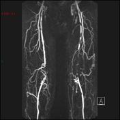
Image 13 MRI ( update )
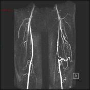
Image 14 MRI ( update )

Image 15 MRI ( update )

Image 16 MRI ( update )


 Unable to process the form. Check for errors and try again.