
Pineal gland
Updates to Article Attributes
The pineal gland is a small, pine-cone shaped structure considered to be part of the epithalamus. It is unpaired and situated in the midline.
Gross anatomy
The pineal gland typically measures 7 x 6 x 3 mm in size and is situated in a groove between the laterally placed thalamic bodies 1-6. Appearing to arise from the gland are two laminae. Above is the habenular commissure and below it is the posterior commissure.
Relations
- anteriorly
-: third ventricle (pineal recess) -
posterioinferiorly -posteroinferiorly: superior cerebellar cistern - superiorly
-: internal cerebral veins, vein of Galen (posteriorly), stria medularis, splenium of the corpus callosum and velum interpositum - inferiorly
-: superior colliculi of the midbrain
Blood supply
The pineal gland receives its blood supply from fine branches off the posterior choroidal arteries and drains superiorly by multiple branches eventually into the great cerebral vein of Galen 3.
Embryology
The pineal gland arises during the seventh week of gestation from a thickening of the ependyma at the posterior most aspect of the third ventricle 5.
Physiology
The pineal gland produces melatonin which affects the modulation of wake/sleep patterns and photoperiodic (seasonal) functions. It is also thought to have a reproductive function and has been associated with the onset of puberty 7. Unlike much of the rest of the brain, it is not isolated from the body by the blood-brain barrier.
Radiographic features
Plain films
The pineal gland has a predilection for calcification which is invariably histologically present in adults but rarely seen below the age of 10 years 6. Calcification is visible on lateral skull x-rays in 50-70% of adults 6. The habenular commissure also calcifies and is visible as a small C-shaped (open part posteriorly) density above and infrontin front of the pineal calcification.
CT
With CT'sCTs far higher contrast resolution calcification is almost always visible in the adult pineal gland, sometimes visible as specs embedded in a small soft tissue nodule similar in density to grey matter. In children under the age of 5 years no calcification is present, but prevalence increases rapidly with age, reaching a plateau at about 30 years of age 5.
MRI
MRI is the modality of choice for evaluating the pineal region although its sensitivity to calcification on conventional sequences is poor (note: susceptibility weighted imaging (SWI) has sensitivity to calcification similar to CT).
The gland appears as a small nodule of tissue with similar intensity to grey matter. It enhances vividly during contrast administration as it is outside the blood brain barrier.
-<li>anteriorly - <a href="/articles/third-ventricle">third ventricle</a> (pineal recess)</li>-<li>posterioinferiorly - <a href="/articles/superior-cerebellar-cistern">superior cerebellar cistern</a>- +<li>anteriorly: <a href="/articles/third-ventricle">third ventricle</a> (pineal recess)</li>
- +<li>posteroinferiorly: <a href="/articles/superior-cerebellar-cistern">superior cerebellar cistern</a>
-<li>superiorly - <a href="/articles/internal-cerebral-vein">internal cerebral veins</a>, <a href="/articles/vein-of-galen">vein of Galen</a> (posteriorly), <a href="/articles/stria-medularis">stria medularis</a>, splenium of the <a href="/articles/corpus-callosum">corpus callosum</a> and <a href="/articles/velum-interpositum">velum interpositum</a>- +<li>superiorly: <a href="/articles/internal-cerebral-vein">internal cerebral veins</a>, <a href="/articles/vein-of-galen">vein of Galen</a> (posteriorly), <a href="/articles/stria-medularis">stria medularis</a>, splenium of the <a href="/articles/corpus-callosum">corpus callosum</a> and <a href="/articles/velum-interpositum">velum interpositum</a>
-<li>inferiorly - superior colliculi of the <a href="/articles/midbrain">midbrain</a>- +<li>inferiorly: superior colliculi of the <a href="/articles/midbrain">midbrain</a>
-</ul><h4>Blood supply</h4><p>The pineal gland receives its blood supply from fine branches off the <a href="/articles/posterior-choroidal-artery">posterior choroidal arteries</a> and drains superiorly by multiple branches eventually into the <a href="/articles/vein-of-galen">great cerebral vein of Galen</a> <sup>3</sup>. </p><h4>Embryology</h4><p>The pineal gland arises during the seventh week of gestation from a thickening of the ependyma at the posterior most aspect of the third ventricle <sup>5</sup>. </p><h4>Physiology</h4><p>The pineal gland produces melatonin which affects the modulation of wake/sleep patterns and photoperiodic (seasonal) functions. It is also thought to have a reproductive function and has been associated with the onset of puberty <sup>7</sup>. Unlike much of the rest of the <a href="/articles/brain">brain</a>, it is not isolated from the body by the <a href="/articles/blood-brain-barrier">blood-brain barrier</a>.</p><h4>Radiographic features</h4><h5>Plain films</h5><p>The pineal gland has a predilection for calcification which is invariably histologically present in adults but rarely seen below the age of 10 years <sup>6</sup>. Calcification is visible on lateral skull x-rays in 50-70% of adults <sup>6</sup>. The habenular commissure also calcifies and is visible as a small C-shaped (open part posteriorly) density above and infront of the pineal calcification. </p><h5>CT</h5><p>With CT's far higher contrast resolution calcification is almost always visible in the adult pineal gland, sometimes visible as specs embedded in a small soft tissue nodule similar in density to grey matter. In children under the age of 5 years no calcification is present, but prevalence increases rapidly with age, reaching a plateau at about 30 years of age <sup>5</sup>. </p><h5>MRI</h5><p>MRI is the modality of choice for evaluating the pineal region although its sensitivity to calcification on conventional sequences is poor (note: <a href="/articles/susceptibility-weighted-imaging">susceptibility weighted imaging (SWI)</a> has sensitivity to calcification similar to CT). </p><p>The gland appears as a small nodule of tissue with similar intensity to grey matter. It enhances vividly during contrast administration as it is outside the <a href="/articles/blood-brain-barrier">blood brain barrier</a>.</p>- +</ul><h4>Blood supply</h4><p>The pineal gland receives its blood supply from fine branches off the <a href="/articles/posterior-choroidal-artery">posterior choroidal arteries</a> and drains superiorly by multiple branches eventually into the <a href="/articles/vein-of-galen">great cerebral vein of Galen</a> <sup>3</sup>. </p><h4>Embryology</h4><p>The pineal gland arises during the seventh week of gestation from a thickening of the ependyma at the posterior most aspect of the third ventricle <sup>5</sup>. </p><h4>Physiology</h4><p>The pineal gland produces melatonin which affects the modulation of wake/sleep patterns and photoperiodic (seasonal) functions. It is also thought to have a reproductive function and has been associated with the onset of puberty <sup>7</sup>. Unlike much of the rest of the <a href="/articles/brain">brain</a>, it is not isolated from the body by the <a href="/articles/blood-brain-barrier">blood-brain barrier</a>.</p><h4>Radiographic features</h4><h5>Plain films</h5><p>The pineal gland has a predilection for calcification which is invariably histologically present in adults but rarely seen below the age of 10 years <sup>6</sup>. Calcification is visible on lateral skull x-rays in 50-70% of adults <sup>6</sup>. The habenular commissure also calcifies and is visible as a small C-shaped (open part posteriorly) density above and in front of the pineal calcification. </p><h5>CT</h5><p>With CTs far higher contrast resolution calcification is almost always visible in the adult pineal gland, sometimes visible as specs embedded in a small soft tissue nodule similar in density to grey matter. In children under the age of 5 years no calcification is present, but prevalence increases rapidly with age, reaching a plateau at about 30 years of age <sup>5</sup>. </p><h5>MRI</h5><p>MRI is the modality of choice for evaluating the pineal region although its sensitivity to calcification on conventional sequences is poor (note: <a href="/articles/susceptibility-weighted-imaging">susceptibility weighted imaging (SWI)</a> has sensitivity to calcification similar to CT). </p><p>The gland appears as a small nodule of tissue with similar intensity to grey matter. It enhances vividly during contrast administration as it is outside the <a href="/articles/blood-brain-barrier">blood brain barrier</a>.</p>
Image 1 Diagram ( update )
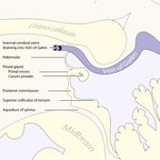
Image 2 MRI (T1) ( update )


 Unable to process the form. Check for errors and try again.