
Spermatic cord hydrocele
Updates to Article Attributes
Body
was changed:
Spermatic cord hydrocele (SCH) refers to loculated fluid collection along the spermatic cord. It separated from and located above the testicle and the epididymis.
Pathology
It results from aberrant closure of the processus vaginalis.
There are two recognised sub types
- encysted hydrocele - fluid collection does not communicate with the peritoneum above or the tunica vaginalis below.
- funicular hydrocele - fluid collection communicates with the peritoneum at the internal inguinal ring but does not communicate with the tunica vaginalis
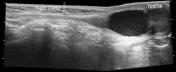
Radiographic features
Ultrasound
Usually seen as an oval anechoic mass in the groin along the spermatic cord, positioned above and separated from the testis and the epididymis. Typically well demarcated. Avascular on colour Doppler interrogation
Treatment and prognosis
Elective surgery may be useful in preventing development of an acquired indirect hernia 3.
Differential diagnosis
It may occasionally be misdiagnosed as an inguinal hernia clinically 7.
See also
- hydrocele
- hydrocele of canal of Nuck: females
-<p><strong>Spermatic cord hydrocele (SCH)</strong> refers to loculated fluid collection along the <a href="/articles/spermatic-cord">spermatic cord</a>. It separated from and located above the testicle and the epididymis. </p><h4>Pathology</h4><p>It results from aberrant closure of the processus vaginalis.</p><p>There are two recognised sub types</p><ul>-<li>encysted hydrocele - fluid collection does not communicate with the peritoneum above or the tunica vaginalis below. </li>- +<p><strong>Spermatic cord hydrocele (SCH)</strong> refers to loculated fluid collection along the <a href="/articles/spermatic-cord">spermatic cord</a>. It separated from and located above the testicle and the epididymis.</p><h4>Pathology</h4><p>It results from aberrant closure of the processus vaginalis.</p><p>There are two recognised sub types</p><ul>
- +<li>encysted hydrocele - fluid collection does not communicate with the peritoneum above or the tunica vaginalis below.</li>
-</ul><h4>Radiographic features</h4><h5>Ultrasound</h5><p>Usually seen as an oval anechoic mass in the groin along the spermatic cord, positioned above and separated from the testis and the epididymis. Typically well demarcated. Avascular on colour Doppler interrogation</p><h4>Treatment and prognosis</h4><p>Elective surgery may be useful in preventing development of an acquired indirect hernia <sup>3</sup>.</p><h4>Differential diagnosis </h4><p>It may occasionally be misdiagnosed as an inguinal hernia clinically<sup> 7</sup>.</p><h4>See also</h4><ul>- +</ul><h4>Radiographic features</h4><h5>Ultrasound</h5><p>Usually seen as an oval anechoic mass in the groin along the spermatic cord, positioned above and separated from the testis and the epididymis. Typically well demarcated. Avascular on colour Doppler interrogation</p><h4>Treatment and prognosis</h4><p>Elective surgery may be useful in preventing development of an acquired indirect hernia <sup>3</sup>.</p><h4>Differential diagnosis</h4><p>It may occasionally be misdiagnosed as an inguinal hernia clinically<sup> 7</sup>.</p><h4>See also</h4><ul>
Images Changes:
Image 3 Ultrasound ( update )

Caption
was changed:
Case 3: encysted
Image 5 Ultrasound (Longitudinal) ( update )
Caption
was changed:
Case5: encysted


 Unable to process the form. Check for errors and try again.