
Sternoclavicular joint series
Updates to Article Attributes
Body
was changed:
The standard SCJsternoclavicular radiographic series is a used to evaluate sternoclavicular joint and the proximal clavicle. Imaging of the sternoclavicular joint has since been replaced by computed tomography.
ProjectionsIndications
Standard projections
Additional projections
Indications
radiographs are performed for a variety of indications including: - trauma
- infection
- deformity in the absence of trauma
- congenital abnormalities
Projections
Standard projections
-
PA view
- standard projection demonstrating the sternoclavicular joints articulating with the manubrium
-
anterior oblique views
- oblique projection best utilised when assessing for joint separation, often performed bi laterally. With the RAO best used to demonstrate the right sternoclavicular joint and the LAO best suited to demonstrate the left sternoclavicular joint
Additional projections
-
lateral sternal view
- radiographic investigation of the entire length of the sternum in profile. The view is used to query fractures or infection
-
serendipity view
- specialised axial projection employed to investigate suspected anterior/posterior dislocations
Related articles
-<p>The standard <strong>SCJ radiographic series</strong> is a used to evaluate <a href="/articles/sternoclavicular-joint">sternoclavicular joint </a>and the proximal <a title="clavicle" href="/articles/clavicle">clavicle</a>.</p><h4>Projections</h4><h5>Standard projections</h5><ul><li><a title="Sternoclavicular joint oblique view" href="/articles/sternoclavicular-joint-oblique-view">Right Anterior Oblique (RAO) + Left Anterior Oblique (LAO)</a></li></ul><h5>Additional projections</h5><ul>-<li><a title="lateral sternal view" href="/articles/lateral-sternal-view">lateral sternal view</a></li>-<li><a title="Sternoclavicular joint PA view" href="/articles/sternoclavicular-joint-pa-view">Sternoclavicular joint PA view</a></li>-</ul><h4>Indications</h4><ul>-<li>trauma</li>- +<p>The <strong>sternoclavicular radiographic series</strong> is a used to evaluate <a href="/articles/sternoclavicular-joint">sternoclavicular joint </a>and the proximal <a href="/articles/clavicle">clavicle</a>. Imaging of the sternoclavicular joint has since been replaced by computed tomography.</p><h4>Indications</h4><p>Sternoclavicular joint radiographs are performed for a variety of indications including: </p><ul>
- +<li>trauma </li>
-<li>deformity in the absence of trauma</li>-<li>congenital abnormalities</li>- +<li>deformity in the absence of trauma </li>
- +<li>congenital abnormalities </li>
- +</ul><h4>Projections</h4><h5>Standard projections</h5><ul>
- +<li>
- +<a title="Sternoclavicular joint (PA view)" href="/articles/sternoclavicular-joint-pa-view">PA view </a><ul><li>standard projection demonstrating the sternoclavicular joints articulating with the manubrium</li></ul>
- +</li>
- +<li>
- +<a href="/articles/anterior-oblique-views">anterior oblique views</a><ul><li>oblique projection best utilised when assessing for joint separation, often performed bi laterally. With the RAO best used to demonstrate the right sternoclavicular joint and the LAO best suited to demonstrate the left sternoclavicular joint </li></ul>
- +</li>
- +</ul><h5>Additional projections</h5><ul>
- +<li>
- +<a href="/articles/sternum-lateral-view-1">lateral sternal view</a><ul><li>radiographic investigation of the entire length of the sternum in profile. The view is used to query <a href="/articles/sternal-fracture">fractures</a> or infection</li></ul>
- +</li>
- +<li>
- +<a href="/articles/serendipity-view">serendipity view</a> <ul><li>specialised axial projection employed to investigate suspected anterior/posterior dislocations </li></ul>
- +</li>
References changed:
- 1. 1. Whitley AS, Sloane C, Hoadley G et-al. Clark's positioning in radiography. Hodder Arnold Publication. ISBN:0340763906. Read it at Google Books - Find it at Amazon
- 2. RT(R) KLBMA, (CT) JLMR. Textbook of Radiographic Positioning and Related Anatomy, 8e. Mosby. ISBN:0323083889. Read it at Google Books - Find it at Amazon
- 3. Bontrager, K.L. and Lampignamo, J.P. Textbook of radiographic positioning and related anatomy. (p. 193–201)6th ed. Mosby, St. Louis, Mo; 2005
- 3. Bontrager, K.L. and Lampignamo, J.P. Textbook of radiographic positioning and related anatomy. (p. 193–201)6th ed. Mosby, St. Louis, Mo; 2005
- 3. 1. Bontrager, K.L. and Lampignamo, J.P. Textbook of radiographic positioning and related anatomy. (p. 193–201)6th ed. Mosby, St. Louis, Mo; 2005
Images Changes:
Image 1 X-ray (Lateral) ( update )
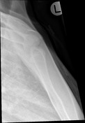
Caption
was changed:
Image 2 X-ray (RAO) ( update )
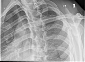
Caption
was changed:
Figure 2: LAO
Image 3 X-ray (LAO) ( update )
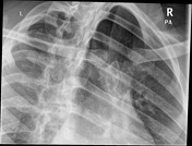
Caption
was changed:
Figure 3: RAO
Image 4 X-ray (serendipity view) ( create )

Image 5 X-ray ( create )


 Unable to process the form. Check for errors and try again.