
Synovial haemangioma
Updates to Article Attributes
Synovial haemangiomas are rare benign vascular malformations that occur in relation to the joint. They are sometimes considered a subtype of soft tissue haemangiomas.
Epidemiology
The lesions typically present in children and young adults.
Clinical presentation
Patients may have pain, swelling and/or limited joint mobility. Occasionally patients can have recurrent haemarthroses 8.
Pathology
The lesions can be cavernous, capillary or mixed.
Location
Most lesions tend to occur around the knee 1.
Radiographic features
Plain radiograph
Plain film findings are generally non-specific and may be seen as a soft tissue mass adjacent to the knee. Accompanying phleboliths may be present.
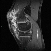
MRI
Typically seen as a lobulated intra-articular mass (although the lesions can be either lobulated or diffuse).
T1: usually of intermediate signal 4
-
T2
markedly hyperintense background; likely from pooled blood within vascular spaces 4
low-signal-intensityhypointense linear structures within the lesion; likely from fibrous septa or vascular channels 4fluid-fluid levels may be present
T1 C+ (Gd): marked enhancement 5
Differential diagnosis
Imaging differential considerations include:
cystic synovial hyperplasia: usually peripheral enhancement only 5
-<p><strong>Synovial haemangiomas</strong> are rare benign vascular malformations that occur in relation to the joint. They are sometimes considered a subtype of <a href="/articles/soft-tissue-venous-malformations">soft tissue haemangiomas</a>.</p><h4>Epidemiology</h4><p>The lesions typically present in children and young adults.</p><h4>Clinical presentation</h4><p>Patients may have pain, swelling and/or limited joint mobility. Occasionally patients can have recurrent haemarthroses <sup>8</sup>.</p><h4>Pathology</h4><p>The lesions can be cavernous, capillary or mixed. </p><h5>Location</h5><p>Most lesions tend to occur around the knee <sup>1</sup>.</p><h4>Radiographic features</h4><h5>Plain radiograph</h5><p>Plain film findings are generally non-specific and may be seen as a soft tissue mass adjacent to the knee. Accompanying phleboliths may be present. </p><h5>MRI</h5><p>Typically seen as a lobulated intra-articular mass (although the lesions can be either lobulated or diffuse).</p><ul>- +<p><strong>Synovial haemangiomas</strong> are rare benign vascular malformations that occur in relation to the joint. They are sometimes considered a subtype of <a href="/articles/soft-tissue-venous-malformations">soft tissue haemangiomas</a>.</p><h4>Epidemiology</h4><p>The lesions typically present in children and young adults.</p><h4>Clinical presentation</h4><p>Patients may have pain, swelling and/or limited joint mobility. Occasionally patients can have recurrent haemarthroses <sup>8</sup>.</p><h4>Pathology</h4><p>The lesions can be cavernous, capillary or mixed. </p><h5>Location</h5><p>Most lesions tend to occur around the knee <sup>1</sup>.</p><h4>Radiographic features</h4><h5>Plain radiograph</h5><p>Plain film findings are generally non-specific and may be seen as a soft tissue mass adjacent to the knee. Accompanying <a href="/articles/phlebolith-1" title="Phlebolith">phleboliths</a> may be present. </p><h5>MRI</h5><p>Typically seen as a lobulated intra-articular mass (although the lesions can be either lobulated or diffuse).</p><ul>
-<li><p>low-signal-intensity linear structures within lesion; likely from fibrous septa or vascular channels <sup>4</sup></p></li>- +<li><p>hypointense linear structures within the lesion; likely from fibrous septa or vascular channels <sup>4</sup></p></li>
-<li><p><strong>T1 C+: </strong>marked enhancement <sup>5</sup></p></li>- +<li><p><strong>T1 C+ (Gd): </strong>marked enhancement <sup>5</sup></p></li>
Image 4 MRI (T1 C+ fat sat) ( update )


 Unable to process the form. Check for errors and try again.