


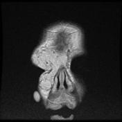


S/S glioma and meningioma... slow progressive loss of vision.
1-optic n. glioma.. ass. with NF1
-<5 ys old
-kincking, buckling of optic nerve
-widening of optoc canal, no hyperostosis.
-no calcification
-bright T2, patchy enhancement.
-iris pigmentation (Lisch nodules)
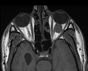
2-optic n. meningioma.... ass. with NF2
-middle age female
-straightening with eccentric thickening of optic n.
- bone hyperostosis, optic canal widening in 10%.
-calcification is com.
-linear band enhancement (tramtrack sign)
-more dense on CT (than glioma)
3-optic neuritis causes: MS, ischemia, vasculitis and Davic syndrome.
4-capillary haemangioma
-grow befor 1y old and involute later.
- no capsule... infiltrate conal and extraconal spaces.
-90%ass. with cutaneous hemangioma.
-MRI :darck T1, bright T2+ curvilinear void signals(vessels)
5-cavernous hemangioma
-adult women.
-has true capsule.
- MRI:similar to CSF+ flow void in the periphery of tumor.
-


 Unable to process the form. Check for errors and try again.