teaching cases
Playlist contributed by
Mohamed Emad Shehata
Play
Share
Playlist
Full screen playlist
Playlist with hidden diagnosis
Full screen playlist with hidden diagnosis
Playlist information
Playlist created:
1 Nov 2020 by
Mohamed Emad Shehata
Last edited:
1 Nov 2020
Number of cases:
201
Number of slides:
0
rID:
134945
Systems:
Breast
,
Cardiac
,
Central Nervous System
,
Chest
,
Forensic
,
Gastrointestinal
,
Gynaecology
,
Head & Neck
,
Hepatobiliary
,
Musculoskeletal
,
Obstetrics
,
Oncology
,
Paediatrics
,
Spine
,
Trauma
,
Urogenital
,
Vascular
Visibility:
public
Show case titles
Case 1
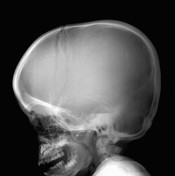
Bilateral Sprengel deformity with Klippel-Feil syndrome
Case 2
Illustration of scurvy signs
Case 3
Illustration of scurvy signs
Case 4
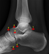
Foot angles - lateral weightbearing foot
Case 5
Shoulder anatomy (illustrations)
Case 6
Illustration of scurvy signs
Case 7
Illustration of scurvy signs
Case 8
Accessory intraparietal suture
Case 9
Diagram - intracranial hemorrhage
Case 10
Ranula
Case 11
Tracheo-esophageal fistula types (diagrams)
Case 12
Middle ear anatomy - annotated CT
Case 13
Inner ear anatomy - annotated CT
Case 14
Normal petrous temporal bone axial CT - with labels
Case 15
How to read a CT of the abdomen and pelvis
Case 16
Pituitary macroadenoma
Case 17
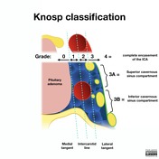
Knosp classification (diagrams)
Case 18
Brain lobes - annotated MRI
Case 19
Cerebral vascular territories (illustration)
Case 20
Common variants of the circle of Willis (illustrations)
Case 21
Central sulcus
Case 22
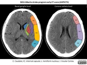
MCA - Alberta stroke program early CT score (ASPECTS) illustration
Case 23
Posterior circulation - Acute stroke prognosis early CT score (pc-ASPECTS) illustration
Case 24
Normal sinus CT (annotated)
Case 25
Teaching head CT with annotated scrollable images
Case 26
Normal sinus CT (annotated)
Case 27
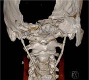
CT neck with annotated scrollable images
Case 28
Normal cervical spine radiographs
Case 29
Ependymoma (cervical cord)
Case 30
Ependymoma (cervical cord)
Case 31
Skull positioning lines (diagram)
Case 32
Jugulotympanic paraganglioma
Case 33
Accessory ossicles of the foot
Case 34
Bone lesion differential diagnosis - illustrations
Case 35
Lisfranc joint - normal alignment
Case 36
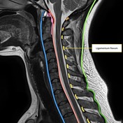
Ligaments of the cervical spine (annotated image)
Case 37
Normal knee MRI
Case 38
Previous breast cancer - axillary surgical clips (chest x-ray)
Case 39
Allergic bronchopulmonary aspergillosis
Case 40
Subclavian steal syndrome
Case 41
Alpha-1-antitrypsin deficiency
Case 42
Centrilobular emphysema
Case 43
Lymphocytic interstitial pneumonia
Case 44
Cystic lung diseases (illustrations)
Case 45
Pulmonary Langerhans cell histiocytosis
Case 46
Neurofibromatosis type 1 - ribbon ribs
Case 47
Amiodarone hepatotoxicity
Case 48
Kerley B lines
Case 49
Upper lobe venous diversion
Case 50
Achalasia
Case 51
Achalasia
Case 52
Achalasia
Case 53
Achalasia
Case 54
Barrett esophagus
Case 55
Esophageal rupture post stricture dilatation
Case 56
Peptic esophageal stricture
Case 57
Normal barium swallow (lateral view)
Case 58
Normal barium swallow - AP pharynx
Case 59
Normal barium swallow (lateral view)
Case 60
Normal small bowel meal
Case 61
How to read a CT of the abdomen and pelvis
Case 62
Gastric bezoar
Case 63
Superior mesenteric artery syndrome
Case 64
Ileocolic intussusception
Case 65
Necrotizing enterocolitis
Case 66
Necrotizing enterocolitis
Case 67
Necrotizing enterocolitis
Case 68
Perforated necrotizing enterocolitis
Case 69
Transient tachypnea of the newborn
Case 70
Meconium peritonitis
Case 71
Cecal volvulus
Case 72
Sigmoid volvulus
Case 73
Frimann-Dahl sign of sigmoid volvulus
Case 74
Endoleak classification (diagram)
Case 75
Renal arterial supply variants (illustrations)
Case 76
Renal angiogram arterial anatomy
Case 77
Pancreas divisum
Case 78
Spleen trauma grading (diagrams)
Case 79
Renal arterial supply variants (illustrations)
Case 80
Aneurysmal bone cyst (ABC)
Case 81
MR angiography of the thoracic aorta
Case 82
Ectopic thoracic kidney
Case 83
Lobster claw sign - papillary necrosis
Case 84
Aortic dissection DeBakey classification (illustration)
Case 85
Thoracic lymph node stations (annotated CT)
Case 86
Achondroplasia
Case 87
Platyspondyly
Case 88
Achondroplasia
Case 89
Bosniak classification of renal cysts (illustrations)
Case 90
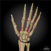
Arterial supply to the hand (illustrations)
Case 91
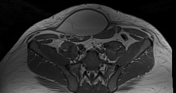
Hilgenreiner's line
Case 92
Hilgenreiner's and Perkin's line (diagram)
Case 93
Acetabular angle - diagram
Case 94
Shenton line
Case 95
Proximal femoral focal deficiency
Case 96
Proximal femoral focal deficiency
Case 97
Innominate artery compression syndrome
Case 98
Hypothenar hammer syndrome
Case 99
Retrocaval (circumcaval) ureter
Case 100
Normal CT abdomen and pelvis: triphasic protocol
Case 101
Mucopolysaccharidoses
Case 102
Metaphyseal corner fracture (child abuse)
Case 103
Metaphyseal corner fracture (child abuse)
Case 104
Cor triatriatum
Case 105
Tuberculous autonephrectomy
Case 106
Tuberculous autonephrectomy
Case 107
Renal tuberculosis with autonephrectomy
Case 108
Renal tuberculosis
Case 109
Paget-Schroetter syndrome
Case 110
Emphysematous pyelitis and pneumoperitoneum
Case 111
Emphysematous pyelitis
Case 112
Emphysematous pyelitis
Case 113
Mesenteric panniculitis and urolithiasis
Case 114
Mesenteric panniculitis and urolithiasis
Case 115
Sigmoid diverticulitis - perforated
Case 116
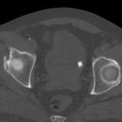
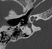
Eagle syndrome
Case 117
Stylohyoid ligament ossification
Case 118
Eagle syndrome
Case 119
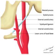
Styloid apparatus (illustration)
Case 120
Long styloid processes/stylohyoid ligaments
Case 121
Right ventricular enlargement on chest radiography (illustration)
Case 122
Right atrial enlargement on chest radiography (illustration)
Case 123
Left atrial enlargement on chest radiography (illustration)
Case 124
Left atrial enlargement on chest radiography (illustration)
Case 125
Left ventricular enlargement on chest radiography (illustration)
Case 126
Cardiomediastinal outlines on chest x-ray
Case 127
Staghorn calculi (coral calculi)
Case 128
Illustration - vesicoureteric reflux grading
Case 129
Vesicoureteric reflux (grade V)
Case 130
Vesicoureteric reflux (grade V)
Case 131
Vesicoureteral reflux
Case 132
Vesicoureteric reflux - grade 4
Case 133
Unilateral vesicoureteric reflux
Case 134
Tetralogy of Fallot
Case 135
Tetralogy of Fallot and tracheoesophageal fistula
Case 136
Tetralogy of Fallot
Case 137
Tetralogy of Fallot with pulmonary stenosis - antenatal
Case 138
Traumatic urethral transection
Case 139
Traumatic urethral transection
Case 140
Mature cystic teratoma
Case 141
Ruptured mature ovarian teratoma
Case 142
Hepatocellular carcinoma
Case 143
Double-layered patella
Case 144
Spinal CSF leak - post-operative (radionuclide cisternography)
Case 145
Jackstone bladder calculus
Case 146
Tricuspid atresia
Case 147
Large skull metastasis: follicular thyroid primary
Case 148
Otospongiosis (fenestral and retrofenestral)
Case 149
Fissula ante fenestram
Case 150
Fenestral otosclerosis
Case 151
Fenestral otosclerosis
Case 152
Bilateral ureterocele with cobra head sign
Case 153
Chiari I and sinus pericranii
Case 154
Sinus pericranii
Case 155
Sinus pericranii
Case 156
Bilateral ovarian fibroma
Case 157
Ovarian fibroma - uterine leiomyomas
Case 158
Transient ischemic dilatation
Case 159
Lymph node levels of the head and neck (annotated CT)
Case 160
Lymph node levels of the head and neck (annotated CT)
Case 161
Perineural tumor spread
Case 162
Tuberculous spondylitis
Case 163
Rectus sheath hematoma
Case 164
Biliary cystadenoma
Case 165
Lymphangioleiomyomatosis
Case 166
Lymphangioleiomyomatosis
Case 167
Le Fort type III fracture
Case 168
Ovarian endometriomas
Case 169
Lipomatous hypertrophy of the interatrial septum
Case 170
Lipomatous hypertrophy of interatrial septum - "cold" on PET-CT
Case 171
Subvalvular aortic stenosis caused by fibrous membrane
Case 172
Aortic valve stenosis
Case 173
Aortic valve stenosis in a case of bicuspid aortic valve
Case 174
Aortic valve stenosis in a case of bicuspid aortic valve
Case 175
Mitral annular calcification
Case 176
Mitral annular calcification
Case 177
Aortic valve stenosis
Case 178
Aortic valve stenosis
Case 179
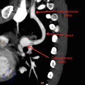
Blalock-Taussig shunt (annotated image)
Case 180
Constrictive pericarditis
Case 181
Pericardial calcification
Case 182
Constrictive pericarditis
Case 183
Benign prostatic hyperplasia
Case 184
Fishhook ureter
Case 185
Prostatomegaly
Case 186
Uterine anatomical abnormalities (illustrations)
Case 187
Total anomalous pulmonary venous return
Case 188
Kümmell disease (avascular necrosis of the vertebrae)
Case 189
Kümmell disease
Case 190
Spinal dermoid
Case 191
Spinal dermoid
Case 192
Retinopathy of prematurity
Case 193
Olivopontocerebellar atrophy
Case 194
Capillary telangiectasia
Case 195
Capillary telangiectasia
Case 196
Capillary telangiectasia
Case 197
Capillary telangiectasia
Case 198
Capillary telangiectasia
Case 199
En plaque meningothelial meningioma with bony inolvement
Case 200
En plaque meningioma - sphenoid wing
Case 201
Atretic parietal cephalocele