
The FRCR 2B rapid-reporting component of the FRCR part 2B is the final part of the FRCR examination. It aims to assess the candidate's ability to accurately detect abnormalities in the type of plain films usually seen in a standard reporting pile.
This is a representative 'packet' of films for this examination. The cases will be viewed in "hidden diagnosis" mode, with the answers available at the end, and following the answers page links to the full cases with ability to view additional views or modalities.


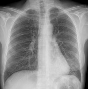


See the full cases by clicking below:
- Case 1: normal hand radiograph (paediatric)
- Case 2: scapholunate interval widening
- Case 3: paraspinal haematoma from three column fracture
- Case 4: lead pipe appearance of ulcerative colitis
- Case 5: normal ankle radiograph
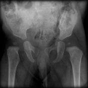
- Case 6: chronic hip subluxation
- Case 7: coin lesion: lung adenocarcinoma
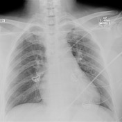
- Case 8: normal thymus on chest radiograph
- Case 9: tibial fracture
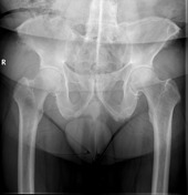
- Case 10: normal pelvic radiograph
- Case 11: normal facial OM30 radiograph
- Case 12: normal lumbar spine radiograph
- Case 13: right retroperitoneal haematoma
- Case 14: normal AP pelvic radiograph
- Case 15: normal PEG projection
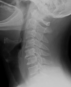
- Case 16: normal lateral soft tissue neck radiograph
- Case 17: bilateral shoulder dislocation on chest radiograph
- Case 18: normal lateral elbow radiograph
- Case 19: neuroblastoma
- Case 20: normal abdominal radiograph
- Case 21: normal skull radiograph
- Case 22: left hilar mass (lung cancer)
- Case 23: ankylosing spondylitis
- Case 24: normal supine chest radiograph
- Case 25: normal shoulder radiograph
- Case 26: perched facet joint
- Case 27: normal chest radiograph (bilateral cervical ribs)
- Case 28: mandibular fracture
- Case 29: left lower lobe pneumonia
- Case 30: normal pelvis radiograph
Want more?
- Radiopaedia.org Rapids practice sets
- Radiopaedia.org's FRCR 2B rapid reporting examination information
- get involved by uploading case to Radiopaedia.org using the tag "rapids"


 Unable to process the form. Check for errors and try again.