
Items tagged “elbow”
136 results found
Case
Supracondylar fracture - marked displacement

Published
20 Jul 2011
94% complete
X-ray
Case
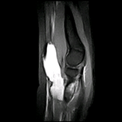
Osteochondritis dissecans of the elbow

Published
24 Aug 2011
45% complete
MRI
Article
Radial head dislocation
Radial head dislocation occurs when the radial head is displaced from its normal articulation with the ulna and the humerus.
The dislocation may be acquired or congenital (see the separate article on congenital radial head dislocation). Additionally, radial head dislocation should be distinguis...
Article
Pulled elbow syndrome
Pulled elbow (also known as nursemaid's elbow) is a subluxation of the radial head into the annular ligament, which usually spontaneously or easily reduces and rarely demonstrates abnormal radiographic features. If the clinical presentation is atypical, pulled elbow should be distinguished from ...
Case
Radial head fracture
Published
03 Jun 2012
94% complete
X-ray
Case
Olecranon fracture

Published
04 Jun 2012
94% complete
X-ray
Case
Radial head fracture
Published
07 Jun 2012
88% complete
Annotated image
X-ray
Article
Elbow
The elbow is a complex synovial joint formed by the articulations of the humerus, the radius, and the ulna.
Gross anatomy
Articulations
The elbow joint is made up of three articulations 2,3:
radiohumeral: capitellum of the humerus with the radial head
ulnohumeral: trochlea of the humerus w...
Article
Flexion supracondylar fracture
Flexion supracondylar humeral fractures account for only 2-4% of all supracondylar fractures 1.
Epidemiology
Unlike the much more common extension supracondylar fracture which are seen in children, flexion fractures are seen in older (adult) patients.
Pathology
They are usually the result of...
Case
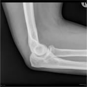
Os supratrochleare dorsale

Published
18 Jun 2012
72% complete
X-ray
Case
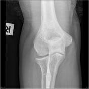
Supracondylar spur

Published
29 Sep 2012
75% complete
X-ray
Case
Olecranon bursitis
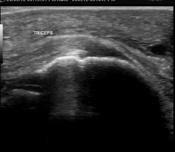
Published
04 Oct 2012
63% complete
Ultrasound
Case
Coronoid process fracture

Published
17 Feb 2013
71% complete
X-ray
Article
Epicondyle fracture (elbow)
Epicondyle fractures are common injuries in children. They represent 10% of all elbow fractures in children and usually occur in boys after a fall on an outstretched arm.
Medial epicondyle fractures comprise most of these injuries. They can usually be treated with splinting and early physiother...
Case
Supracondylar fracture

Published
25 Mar 2013
66% complete
X-ray
Article
Anterior humeral line
The anterior humeral line is key to demonstrating normal elbow alignment and should be used whenever reading a pediatric elbow radiograph to exclude a subtle supracondylar fracture.
Measurement
A line drawn down the anterior surface of the humerus should intersect the middle third of the capit...
Article
Radiocapitellar line
The radiocapitellar line is one of the key lines used to assess alignment on the elbow radiograph. It is particularly useful in the pediatric setting.
Measurement
A line drawn down the neck of the radius should intersect the capitellum. It is important to ensure that you draw the line down the...
Case
Supracondylar fracture - grade Ib

Published
06 Apr 2013
88% complete
Annotated image
X-ray
Case
Supracondylar fracture

Published
06 Apr 2013
72% complete
X-ray
Case
Bicipital radial bursitis
Published
25 Apr 2013
80% complete
MRI


 Unable to process the form. Check for errors and try again.