
Items tagged “pocus”
122 results found
Case
Apical 4 chamber view - normal (transthoracic echocardiography)

Published
28 Aug 2018
47% complete
Ultrasound
Article
Focus‐assessed transthoracic echocardiography
FATE (focus‐assessed transthoracic echocardiography) is a goal-directed protocol used in critical care for indications such as hemodynamic instability, shock, and pulseless electrical activity (PEA) arrest 1.
The protocol is designed as a series of questions as follows:
does the left ventri...
Case
Normal trachea (ultrasound)

Published
29 Aug 2018
25% complete
Ultrasound
Case
Small bowel (ultrasound)

Published
30 Sep 2018
60% complete
Ultrasound
Case
Thoracic aorta - normal (transthoracic echocardiography)

Published
29 Aug 2018
50% complete
Ultrasound
Article
Point-of-care ultrasound (curriculum)
The point-of-care ultrasound (PoCUS) curriculum is one of our curriculum articles and aims to be a collection of articles that represent the core applications of ultrasonography in a point-of-care setting.
Point-of-care ultrasound refers to ultrasonography which may be simultaneously performed,...
Article
Interscalene brachial plexus block
An interscalene brachial plexus block is indicated for procedures involving the shoulder and upper arm.
History
Ultrasound-guided brachial plexus nerve blocks entered the literature in 1989, when Ting et al. detailed their success with axillary nerve blocks in 10 patients 3.
Indications
r...
Article
Left ventricular ejection fraction (echocardiography)
Left ventricular ejection fraction (LVEF) is a surrogate for left ventricular global systolic function, defined as the left ventricular stroke volume divided by the end-diastolic volume.
Terminology
Point-of-care echocardiography protocols typically use a semi-quantitative approach in defining...
Case
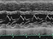
Mitral valve (M-mode echocardiogram)
Published
25 Sep 2018
57% complete
Ultrasound
Article
Carpentier classification of mitral valve regurgitation
The Carpentier classification divides mitral valve regurgitation into three types based on leaflet motion 1:
type I: normal leaflet motion
annular dilation, leaflet perforation
regurgitation jet directed centrally
type II: excessive leaflet motion
papillary muscle rupture, chordal rupture, ...
Article
Right ventricular function (point of care ultrasound)
Right ventricular function is often measured in point-of-care ultrasonography as a composite of the right ventricular size, wall measurements, and contractile efforts.
Terminology
The right ventricle (RV) can be anatomically divided into an inflow portion, an outflow portion, and an apex. Con...
Case
Aortic stenosis (transthoracic echocardiography)
Published
30 Sep 2018
82% complete
Ultrasound
Article
A-line (ultrasound)
An A-line is an ultrasonographic artifact appreciated during the insonation of an aerated lung. 1
The term may be applied to the horizontal, echogenic long path reverberation artifacts that occur beneath the pleural line at multiples of the distance between the ultrasound probe and the visceral...
Article
Pleural effusion volume (ultrasound)
Measurement of a pleural effusion volume with point-of-care ultrasonography may be a useful tool for intensivists and is an active area of research in critical care 7.
In controlled settings ultrasound may detect constitutive pleural fluid, can reliably detect effusions >20 mL in clinical setti...
Case
Pleural effusion (ultrasound)

Published
01 Oct 2018
72% complete
Ultrasound
Article
Raised intracranial pressure
Raised intracranial pressure is a pathological increase in the intracranial pressure and is a medical emergency.
Clinical presentation
The symptoms and signs of raised intracranial pressure are often non-specific and insidious in onset:
headache
drowsiness
anorexia
visual disturbances
bl...
Article
Beads on a string sign (chronic salpingitis)
The beads on a string sign is used to refer to the classic ultrasound morphologic changes of the fallopian tubes as a result of chronic salpingitis.
Terminology
The "string" alludes to the notably thin salpingeal wall, while the hyperechoic mural nodules constitute the "beads" 1.
Pathology...
Case
Tricuspid regurgitation (echocardiography)

Published
14 Nov 2018
72% complete
Ultrasound
Case
FAST exam - normal (ultrasonography)

Published
16 Nov 2018
60% complete
Ultrasound
Article
B-line (ultrasound)
The B-line is an artifact relevant in lung ultrasonography. As originally described, it has seven defining features 1:
a hydroaeric comet-tail artifact
arising from the pleural line
hyperechoic
well-defined
extending indefinitely
erasing A-lines
moving in concert with lung sliding, if lung...


 Unable to process the form. Check for errors and try again.