
Items tagged “spinal cord”
26 results found
Article
Hydromyelia
In hydromyelia, there is dilatation of the central canal of the spinal cord. The dilatation is lined by the normal ependymal lining of the central canal.
The term can refer to dilatation of the persistent central canal of the spinal cord which communicates with the fourth ventricle (cavity wall...
Case
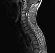
Diastematomyelia

Published
24 Apr 2010
92% complete
MRI
Case
Arachnoid cyst - spinal

Published
16 Jun 2010
71% complete
MRI
Case
Von Hippel-Lindau disease with spinal haemangioblastoma
Published
17 Sep 2010
71% complete
MRI
Ultrasound
Case
Intradural spinal lipoma

Published
07 May 2011
92% complete
MRI
Case
Spinal cord metastasis

Published
13 May 2011
80% complete
MRI
Case
Syringomyelia

Published
26 Jun 2011
53% complete
MRI
Case
Spinal cord ischaemia

Published
17 Mar 2016
91% complete
MRI
CT
Case
Spinal cord ischaemia

Published
06 Jun 2016
95% complete
MRI
CT
Case
Filar cyst

Published
02 Aug 2016
57% complete
Ultrasound
Article
Anterior corticospinal tract
The anterior corticospinal tract is formed at the level of the of the medullary pyramids, where the majority (90%) of descending corticospinal tract fibres decussate to form the lateral corticospinal tract. The majority of the remaining non-decussating 10% of fibres form the much smaller anterio...
Article
Lateral corticospinal tract
The lateral corticospinal tract is formed at the level of the of the medullary pyramids when the majority (90%) of descending corticospinal tract fibres decussate. The remaining 10% do not decussate and form the much smaller anterior corticospinal tract. A few non-decussated fibres may enter the...
Article
Spinocerebellar tract
The spinocerebellar tracts are afferent neurones that convey proprioceptive data from the spinal cord to the cerebellum. There are anterior and posterior spinocerebellar tracts, also eponymously named the Gowers tract and Flechsig tract respectively. Both the anterior and posterior spinocerebell...
Article
Corticorubral tract
The corticorubral tract contains neurones that connect the primary motor and sensory areas to the red nucleus. The rubrospinal tract then descends through the spinal cord.
The tract is thought to excite flexor muscles and inhibit extensor muscles.
Gross anatomy
Central connections
The cort...
Article
Rubrospinal tract
The rubrospinal tract contains neurones that carry signals from the corticorubral tract. The tract is thought to excite flexor muscles and inhibit extensor muscles.
Gross anatomy
Central connections
The magnocellular portion of the red nucleus gives rise to the rubrospinal tract. It decussate...
Article
Lateral spinothalamic tract
The lateral spinothalamic tract, also known as the lateral spinothalamic fasciculus, is an ascending pathway located anterolaterally within the peripheral white matter of the spinal cord. It is primarily responsible for transmitting pain and temperature as well as coarse touch.
The anterior sp...
Article
Anterior spinothalamic tract
The anterior spinothalamic tract, also known as the ventral spinothalamic fasciculus, is an ascending pathway located anteriorly within the spinal cord, primarily responsible for transmitting coarse touch and pressure.
The lateral spinothalamic tract (discussed separately), in contrast, primar...
Case
Dorsal thoracic arachnoid web

Published
18 Nov 2016
32% complete
Diagram
Case
Spinal cavernoma

Published
19 Jul 2017
68% complete
MRI
Article
Denticulate ligaments
The denticulate ligaments are bilateral triangular lateral extensions of pia mater that anchor the spinal cord to the dura mater.
They are formed by pia mater of the spinal cord coursing in-between the dorsal and ventral nerve roots bilaterally. They function to provide stability to the spinal ...


 Unable to process the form. Check for errors and try again.