
Items tagged “tumour”
73 results found
Case
Diffuse astrocytoma NOS ("protoplasmic")

Published
08 Jul 2013
92% complete
MRI
Case
Supratentorial pilocytic astrocytoma

Published
08 Jul 2013
95% complete
MRI
Case
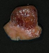
Non-seminomatous germ cell tumor - testis (gross pathology)

Published
06 Jan 2014
54% complete
Pathology
Case
Papillary renal cell carcinoma (pathology)
Published
06 Feb 2014
88% complete
Pathology
Case
Ovarian germ cell tumor

Published
16 Feb 2014
74% complete
Ultrasound
X-ray
CT
Article
Intracranial tumors (summary)
This is a basic article for medical students and other non-radiologists
Intracranial tumors comprise a heterogeneous group of tumors. In adult patients, the majority represent metastatic disease with a smaller proportion being primary brain tumors. Metastasis to the brain occurs, most commonly,...
Case
Glioblastoma NOS

Published
14 Jun 2015
98% complete
CT
MRI
Case
Ecchordosis physaliphora

Published
05 Jun 2018
82% complete
CT
MRI
Case
Hypothalamic germinoma

Published
24 Jan 2018
71% complete
MRI
Case
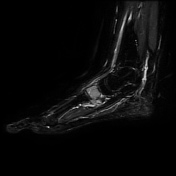
Chondroblastoma - midfoot
Published
11 Dec 2018
77% complete
X-ray
MRI
Article
Half-moon sign (femoral neck)
The half-moon sign describes the morphology of bone marrow edema at the femoral neck on fluid-sensitive MRI sequences, which can be seen in osteoid osteoma or stress fractures 1-3.
Differential diagnosis
intra-articular osteoid osteoma
in patients without a history of overuse, it is highly s...
Article
Apical chest mass
Apical chest masses are often important and may be missed, especially when examined with a plain chest radiograph. It is always recommended to perform a targeted assessment of the apices of the lungs during a chest x-ray; they are one of the classic review areas.
Pathology
Etiology
Commonly a...
Article
Central scar in hepatic lesions
The central scar in hepatic lesions most frequently has been described in focal nodular hyperplasia which the scar is T2 hyperintense and usually non-calcified, and fibrolamellar hepatocellular carcinoma, where the scar is T2 hypointense and often calcified. Scars do not have to be exactly centr...


 Unable to process the form. Check for errors and try again.