


213 results found
Case
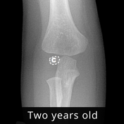
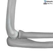
Elbow ossification centers
Published
17 Apr 2024
41% complete
Annotated image
Case
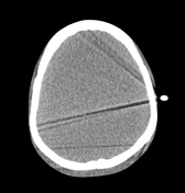
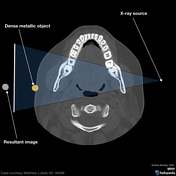
Streak artefact due to leads - anthropomorphic phantom (CT head)
Published
16 Apr 2024
47% complete
CT
Case
Apophyseal avulsion fractures of the pelvis and hip (illustration)

Published
01 Apr 2024
44% complete
Diagram
Case
Blood supply of the femoral head (illustration)

Published
05 Feb 2024
44% complete
Diagram
Case
Slipped capital femoral epiphysis (illustrations)

Published
26 Dec 2023
41% complete
Diagram
Case
Rectal anatomy (illustrations)

Published
24 May 2023
41% complete
Diagram
Case
Pediatric hand anatomy (illustration)

Published
24 May 2023
35% complete
Diagram
Case
Dislodgement of PICC after contrast injection

Published
21 Mar 2023
83% complete
CT
Case
Dislodgement of PICC after contrast injection

Published
21 Mar 2023
87% complete
Fluoroscopy
CT
Case
Lauge-Hansen classification of ankle fractures (illustrations)

Published
01 Nov 2022
36% complete
Diagram
Case
Ankle tear drop sign (illustration)

Published
01 Nov 2022
41% complete
Diagram
Case
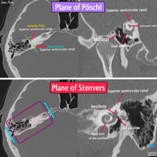
Temporal bone CT planes
Published
31 Aug 2022
42% complete
Annotated image
CT
Case
Chopart and Lisfranc joints (illustrations)

Published
29 Aug 2022
41% complete
Diagram
Case
Carpal dislocations (illustrations)

Published
09 Aug 2022
38% complete
Diagram
Case
Nunley-Vertullo classification of Lisfranc injuries (illustrations)

Published
04 May 2022
41% complete
Diagram
Case
Lisfranc injury - Myerson classifications (illustrations)

Published
04 May 2022
35% complete
Diagram
Case
Lisfranc ligamentous complex (illustration)

Published
03 May 2022
44% complete
Diagram
Case
Regions of the foot (illustrations)

Published
02 May 2022
44% complete
Diagram
Case
Accessory ossicles of the foot (illustration)

Published
02 May 2022
44% complete
Diagram
Case
Elbow anatomy (illustration)

Published
18 Apr 2022
35% complete
Diagram
Case
Supracondylar fracture (illustrations)

Published
14 Feb 2022
35% complete
Diagram
Case
Pediatric elbow anatomy (illustrations)

Published
08 Feb 2022
35% complete
Diagram
Case
Milch classification of lateral humeral condyle fractures (illustrations)

Published
08 Feb 2022
44% complete
Diagram
Case
Hindfoot alignment view

Published
10 Jan 2022
44% complete
Annotated image
Case
Clements-Nakayama positioning

Published
06 Dec 2021
47% complete
Annotated image
Case
Lateral knee positioning

Published
28 Oct 2021
44% complete
Annotated image
Case
Patterns of knee injury

Published
18 Oct 2021
41% complete
Diagram
Case
Knee joint dislocations (illustrations)

Published
17 Oct 2021
36% complete
Diagram
Case
AO classification of distal femur fractures (illustration)
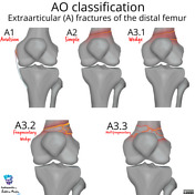
Published
17 Oct 2021
44% complete
Diagram
Case
Schatzker classification of tibial plateau fractures (illustrations)

Published
09 Oct 2021
35% complete
Diagram
Case
Knee radiograph checklist (illustration)

Published
08 Oct 2021
44% complete
Diagram
Case
Lipohemarthrosis and hemarthrosis (illustrations)

Published
08 Oct 2021
44% complete
Diagram
Case
Knee anatomy (illustrations)

Published
04 Oct 2021
35% complete
Diagram
Case
Mallet finger (illustration)

Published
22 Apr 2021
41% complete
Diagram
Case
Elbow joint effusion (illustration)
Published
08 Feb 2021
35% complete
Diagram
Case
AO/OTA classification of distal humeral fractures

Published
15 Jan 2021
35% complete
Diagram
Case
Scapholunate advanced collapse (illustration)

Published
14 Aug 2020
32% complete
Diagram
Case
Normal wrist alignment

Published
13 Aug 2020
38% complete
Case
Scaphoid non-union advanced collapse (illustration)

Published
12 Aug 2020
32% complete
Diagram
Case
Radial height (illustration)

Published
11 Aug 2020
41% complete
Diagram
Case
Normal wrist alignment, dorsal and volar intercalated segmental instability (illustration)

Published
10 Aug 2020
35% complete
Diagram
Case
Ossicles of the wrist and hand (illustration)
Published
10 Aug 2020
29% complete
Diagram
Case
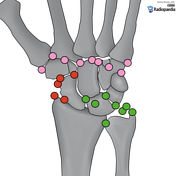
Wrist anatomy (illustration)

Published
10 Aug 2020
29% complete
Diagram
Case
Lateral wrist anatomy (illustration)

Published
09 Aug 2020
35% complete
Diagram
Case
Ghost image (diagram)
Published
09 Apr 2020
35% complete
Case
Rockwood classification of acromioclavicular joint injury

Published
25 Nov 2019
22% complete
Diagram
Case
Shoulder series

Published
22 Nov 2019
41% complete
X-ray
Case
Images for MCQs

Published
08 Oct 2019
18% complete
Diagram
Case
Recurrent neural network

Published
05 Jul 2019
22% complete
Diagram
Case
Simplified neural network

Published
09 Jun 2019
35% complete
Diagram
Case
Hip views

Published
06 Jun 2019
19% complete
Case
Hip series (trauma)

Published
06 Jun 2019
38% complete
Case
Hip series

Published
06 Jun 2019
22% complete
Case
Femur series

Published
06 Jun 2019
25% complete
Annotated image
Case
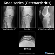
Knee series (osteoarthritis)
Published
06 Jun 2019
41% complete
Annotated image
Case
Knee series

Published
06 Jun 2019
22% complete
Case
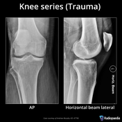
Knee series (trauma)
Published
06 Jun 2019
44% complete
Annotated image
Case
Tibia and fibula series

Published
06 Jun 2019
41% complete
Annotated image
Case
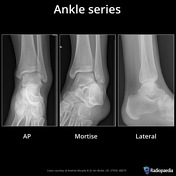
Ankle series
Published
05 Jun 2019
22% complete
Annotated image
Case
Foot series

Published
04 Jun 2019
22% complete
Annotated image
Case
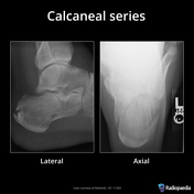
Calcaneal series
Published
04 Jun 2019
35% complete
Annotated image
Case
Convolutional neural network (diagram)

Published
01 May 2019
44% complete
Diagram
Case
TS-OP line (diagram)

Published
02 Oct 2018
25% complete
Diagram
Case
Grid cut off - upside down grid

Published
20 Aug 2018
85% complete
X-ray
Case
Anterior shoulder dislocation

Published
12 Aug 2018
75% complete
X-ray
Case
Hill-Sachs lesion - chronic

Published
09 Aug 2018
91% complete
X-ray
Case
Thomas Edison (diagram)

Published
06 Jul 2018
35% complete
Photo
Case
Clothing artifact

Published
26 Apr 2018
91% complete
X-ray
Case
Photographic plate made by Henri Becquerel (photo)

Published
21 Mar 2018
32% complete
Photo
Case
Antoine Henri Becquerel (photo)

Published
21 Mar 2018
38% complete
Photo
Case
Skull positioning lines (diagram)

Published
03 Mar 2018
38% complete
Diagram
Case
Cranial landmarks (photo)

Published
03 Mar 2018
38% complete
Annotated image
Case
Cervical spine anatomy

Published
03 Mar 2018
44% complete
Annotated image
Case
Marie Curie (photo)

Published
22 Feb 2018
29% complete
Case
Hounsfield's sketch (photo)

Published
05 Feb 2018
29% complete
Photo
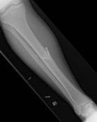
Case
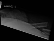
Bilateral femoral fractures (Clements-Nakayama view)
Published
01 Oct 2017
85% complete
X-ray
Case
Pectoralis major tear

Published
27 Sep 2017
89% complete
MRI
Case
Carpal fractures - smart watch impaction

Published
27 Sep 2017
92% complete
X-ray
CT
Case
Malpositioned nasogastric tube

Published
12 Sep 2017
77% complete
X-ray
Annotated image
Case
Tiger tube

Published
11 Sep 2017
52% complete
Annotated image
X-ray
Case
Contrast extravasation

Published
11 Sep 2017
88% complete
X-ray
Case
Stenvers view

Published
11 Sep 2017
44% complete
X-ray
Case
Contrast extravasation

Published
11 Sep 2017
79% complete
X-ray
Case
Plombage (Lucite spheres)

Published
11 Sep 2017
91% complete
X-ray
Case
Contrast staining post endovascular clot retrieval
Published
11 Sep 2017
83% complete
CT
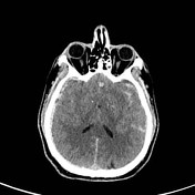
Case
Hemorrhagic transformation of left middle cerebral artery territory infarct

Published
09 Sep 2017
89% complete
CT
Case
Failed intraosseous contrast injection

Published
09 Sep 2017
89% complete
CT
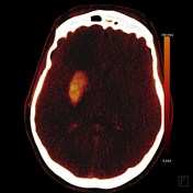
Case
Traumatic brain injury (intraosseous vascular access)
Published
09 Sep 2017
86% complete
CT
Nuclear medicine
Case
Neck CT angiogram (intraosseous vascular access)

Published
09 Sep 2017
89% complete
CT
Case
Sigmoid volvulus

Published
05 Sep 2017
92% complete
X-ray
CT
Case
Breast abscess

Published
31 Aug 2017
66% complete
Ultrasound
X-ray
Case
Foreign body (chronic)
Published
30 Aug 2017
88% complete
X-ray
Case
Cassette artifact

Published
30 Aug 2017
94% complete
X-ray
Case
Pubic symphysis disruption

Published
26 Aug 2017
88% complete
X-ray
ADVERTISEMENT: Supporters see fewer/no ads


 Unable to process the form. Check for errors and try again.