
Abdominal aortic aneurysm (summary)
Updates to Article Attributes
Abdominal aortic aneurysms (AAA) are focal dilatations of the abdominal aorta that are 50% greater than the proximal normal segment or that are >3 cm in maximum diameter.
Read more: abdominal aortic aneurysm
Summary
-
epidemiology
- prevalence of rupture increases with age
- males more commonly affected than females
- almost 10% of people over 65 have an aneurysm
- 10 commonest cause of death in Western populations
-
presentation
- may be asymptomtic
- pain if rapid change in diameter or impending rupture
- pain and shock in rupture
-
pathology
- many processes may cause aneurysms
- atherosclerosis is by far the commonest cause
- inflammatory, infective and vasculitic conditions may also be causes
- many processes may cause aneurysms
-
radiology
- US great for population screening and monitoring small aneurysms
- CT is the gold-standard test for aneurysm assessment
- CT is used in the acute setting of potential aneurysm complciation
-
treatment
- small aneurysms without signs of complication are followed up
-
the larger the aneurysm the more likely it is to rupture
- aneurysmal rupture carries a significant risk of death
- larger, complicated aneurysms
can be stentedneed treatment- endovascular (EVAR) or open surgery can be performed
-
prognosis-
the larger the aneurysm the more likely it is to ruptureaneurysmal rupture carries a significant mortality risk
-
Radiographic features
Role of imaging
- detection of AAA
- monitoring of the rate of growth
- pre-operative planning
- post-operative follow-up
Plain radiograph
An aneurysm may be visible as an area of curvilinear calcification in the paravertebral region on either abdominal or lumbar spine radiographs performed for alternative indications.
Ultrasound
Ultrasound assessment is simple, safe and inexpensive. It has a reported sensitivity of 95% and specificity close to 100%. It is usually the preferred choice for monitoring small aneurysms.
CT
CT angiography is considered the imaging gold standard but has a high radiation dose. Excellent for pre-operative planning as it accurately delineates the size and shape of the AAA and its relationship to branch arteries and the aortic bifurcation.
As aneurysms increase in size the risk of complications increase. CT can be used to make an assessment of rupture, impending rupture or contained leak.
Diameter increase by 10 mm over 12 months, or a diameter of 7 cm are taken to be at high risk for rupture and may warrant urgent repair.
Read more
-<p><strong>Abdominal aortic aneurysms (AAA)</strong> are focal dilatations of the abdominal aorta that are 50% greater than the proximal normal segment or that are >3 cm in maximum diameter.</p><p>Read more: <a title="Abdominal aortic aneurysm" href="/articles/abdominal-aortic-aneurysm">abdominal aortic aneurysm</a></p><h4>Summary</h4><ul>- +<p><strong>Abdominal aortic aneurysms (AAA)</strong> are focal dilatations of the abdominal aorta that are 50% greater than the proximal normal segment or that are >3 cm in maximum diameter.</p><h4>Summary</h4><ul>
-<li>larger, complicated aneurysms can be stented<ul><li>endovascular (EVAR) or open surgery can be performed</li></ul>- +<li>the larger the aneurysm the more likely it is to rupture<ul><li>aneurysmal rupture carries a significant risk of death</li></ul>
-</ul>- +<li>larger, complicated aneurysms need treatment<ul><li>endovascular (EVAR) or open surgery can be performed</li></ul>
-<li>-<strong>prognosis</strong><ul><li>the larger the aneurysm the more likely it is to rupture<ul><li>aneurysmal rupture carries a significant mortality risk</li></ul>-</li></ul>- +</ul>
-</ul><h5>Plain radiograph</h5><p>An aneurysm may be visible as an area of curvilinear calcification in the paravertebral region on either abdominal or lumbar spine radiographs performed for alternative indications.</p><h5>Ultrasound</h5><p>Ultrasound assessment is simple, safe and inexpensive. It has a reported sensitivity of 95% and specificity close to 100%. It is usually the preferred choice for monitoring small aneurysms.</p><h5>CT </h5><p>CT angiography is considered the imaging gold standard but has a high radiation dose. Excellent for pre-operative planning as it accurately delineates the size and shape of the AAA and its relationship to branch arteries and the aortic bifurcation.</p><p>As aneurysms increase in size the risk of complications increase. CT can be used to make an assessment of rupture, impending rupture or contained leak.</p><p>Diameter increase by 10 mm over 12 months, or a diameter of 7 cm are taken to be at high risk for rupture and may warrant urgent repair.</p>- +</ul><h5>Plain radiograph</h5><p>An aneurysm may be visible as an area of curvilinear calcification in the paravertebral region on either abdominal or lumbar spine radiographs performed for alternative indications.</p><h5>Ultrasound</h5><p>Ultrasound assessment is simple, safe and inexpensive. It has a reported sensitivity of 95% and specificity close to 100%. It is usually the preferred choice for monitoring small aneurysms.</p><h5>CT </h5><p>CT angiography is considered the imaging gold standard but has a high radiation dose. Excellent for pre-operative planning as it accurately delineates the size and shape of the AAA and its relationship to branch arteries and the aortic bifurcation.</p><p>As aneurysms increase in size the risk of complications increase. CT can be used to make an assessment of rupture, impending rupture or contained leak.</p><p>Diameter increase by 10 mm over 12 months, or a diameter of 7 cm are taken to be at high risk for rupture and may warrant urgent repair.</p><h4>Read more</h4><ul><li><a href="/articles/abdominal-aortic-aneurysm">abdominal aortic aneurysm</a></li></ul>
Tags changed:
- summary
- medical student
Systems changed:
- Vascular
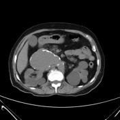
Image 1 CT (C+ portal venous phase) ( create )

Image 2 CT (C+ arterial phase) ( create )
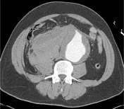
Image 3 CT (C+ portal venous phase) ( create )
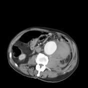
Image 4 CT (non-contrast) ( create )


 Unable to process the form. Check for errors and try again.