
Acute cerebellitis
Updates to Article Attributes
Acute cerebellitis (AC), also known as acute cerebellar ataxia, is a rare inflammatory process characterised by a sudden onset of cerebellar dysfunction usually affecting children. It is related as a consequence of a primary or secondary infection, or as a result of post-vaccinal reaction.
Epidemiology
Usually occurring in children under six years of age, AC is the most common cause of ataxia in the paediatric population. It is reported as a complication of several systemic infectious diseases, such as Varicella (chickenpox) 6.
Adult cases of cerebellitis are possible and well established in medical literature 5.
Clinical presentation
A broad range of signs and symptoms may be present, such as: fever, tremor, nystagmus, truncal ataxia, dysarthria, headache, nausea, vomiting and consciousness alterations. Signs of meningeal irritation and seizures may be observed less frequently 1,4.
Some relevant complications have been described, such as:
- obstructive hydrocephalus and consequent intracranial hypertension
- tonsillar herniation
- cerebellar trunk compression
- severe cerebellar atrophy 3
Laboratory findings:
CSF pleocytosis 4
Pathology
Sometimes the etiologic agentAC has been associated with a large number of the AC cannot be isolatedinfectious agents, such as: coxsackievirus, echovirus, enteroviruses, Epstein-Barr virus, hepatitis A, herpes simplex virus I, human herpesvirus 6, measles, rubella, mumps, parvovirus B19, Borrelia burgdorferi (Lyme disease), malaria, mycoplasma pneumoniae, and typhoid fever 2,4,6.
Acute cerebellar ataxia following vaccination for varicella, hepatitis B, and rabies were reported 6-8.
Radiographic features
CT
Cerebellar images can be normal due the CT limitations on the evaluation of posterior fossa. Nonetheless, complications like compression of the brain stem and obstructive hydrocephalus, when present, are identified on CT images and may guide for further investigation.
MRI
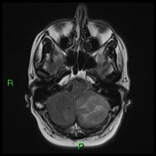
Usually the cerebellar hemispheres are symmetric. It may show a homogeneous mass effect due the inflammatory processcerebellar swelling 1,4.
- T1:cortical hypointensity
- T2/FLAIR: cortical hyperintensity
- DWI/ADC: may show restriction due a cytotoxic oedema caused by the acute inflammatory lesions 4
- T1 C+ (Gd): cortical and adjacent leptomeninges may show enhancement
Treatment and prognosis
The disease is generally benign and self-limited, however some complications, as described above, can take the patient to a worse prognosis and lead to death.
In rare complicated cases a massive cerebellar oedema can require a surgical decompression of the posterior cranial fossa 4.
Differential diagnosis
Clinical features and the age group are essential when thinking in possible differential diagnosis. AC limited to one cerebellar hemisphere only may mimic a cerebellar tumour, especially:
- Lhermitte-Duclos disease
- Cerebellar lymphoma
-<p><strong>Acute cerebellitis (AC)</strong>, also known as <strong>acute cerebellar ataxia</strong>, is a rare inflammatory process characterised by a sudden onset of cerebellar dysfunction usually affecting children. It is related as a consequence of a primary or secondary infection, or as a result of post-vaccinal reaction.</p><h4>Epidemiology</h4><h4>Clinical presentation</h4><p>A broad range of signs and symptoms may be present, such as: fever, tremor, nystagmus, truncal ataxia, dysarthria, headache, nausea, vomiting and consciousness alterations. Signs of meningeal irritation and seizures may be observed less frequently <sup>1,4</sup>. </p><p>Some relevant complications have been described, such as:</p><ul>-<li>obstructive hydrocephalus and consequent intracranial hypertension </li>-<li>tonsillar herniation </li>- +<p><strong>Acute cerebellitis (AC)</strong>, also known as <strong>acute cerebellar ataxia</strong>, is a rare inflammatory process characterised by a sudden onset of cerebellar dysfunction usually affecting children. It is related as a consequence of a primary or secondary infection, or as a result of post-vaccinal reaction.</p><h4>Epidemiology</h4><p>Usually occurring in children under six years of age, AC is the most common cause of ataxia in the paediatric population. It is reported as a complication of several systemic infectious diseases, such as Varicella (chickenpox) <sup>6</sup>. </p><p>Adult cases of cerebellitis are possible and well established in medical literature <sup>5</sup>. </p><h4>Clinical presentation</h4><p>A broad range of signs and symptoms may be present, such as: fever, tremor, nystagmus, truncal ataxia, dysarthria, headache, nausea, vomiting and consciousness alterations. Signs of meningeal irritation and seizures may be observed less frequently <sup>1,4</sup>. </p><p>Some relevant complications have been described, such as:</p><ul>
- +<li>
- +<a title="Obstructive hydrocephalus" href="/articles/obstructive-hydrocephalus">obstructive hydrocephalus</a> and consequent intracranial hypertension </li>
- +<li><a title="Cerebral herniation" href="/articles/cerebral-herniation">tonsillar herniation </a></li>
-<li>severe cerebellar atrophy <sup>3</sup>- +<li>severe <a title="Diffuse cerebellar atrophy" href="/articles/diffuse-cerebellar-atrophy">cerebellar atrophy</a> <sup>3</sup>
-</ul><h4>Pathology</h4><p><br>Sometimes the etiologic agent of the AC cannot be isolated. </p><h4>Radiographic features</h4><h5>CT</h5><p>Cerebellar images can be normal due the CT limitations on the evaluation of posterior fossa. Nonetheless, complications like obstructive hydrocephalus, when present, are identified on CT images and may guide for further investigation. </p><h5>MRI </h5><p>Usually the cerebellar hemispheres are symmetric. It may show a homogeneous mass effect due the inflammatory process. <br><strong>T1:</strong> cortical hypointensity <br><strong>T2/FLAIR:</strong> cortical hyperintensity <br><strong>T1 C+ (Gd):</strong> cortical and adjacent leptomeninges may show enhancement </p><h4>Treatment and prognosis</h4><p>The disease is generally benign and self-limited, however some complications, as described above, can take the patient to a worse prognosis and lead to death. </p>- +</ul><h6>Laboratory findings:</h6><ul><li><p>CSF pleocytosis <sup>4</sup> </p></li></ul><h4>Pathology</h4><p>AC has been associated with a large number of infectious agents, such as: coxsackievirus, echovirus, enteroviruses, Epstein-Barr virus, hepatitis A, herpes simplex virus I, human herpesvirus 6, measles, rubella, mumps, parvovirus B19, <em>Borrelia burgdorferi</em> (Lyme disease), malaria, <em>mycoplasma pneumoniae</em>, and typhoid fever <sup>2,4,6</sup>.</p><p>Acute cerebellar ataxia following vaccination for varicella, hepatitis B, and rabies were reported <sup>6-8</sup>. </p><h4><strong>Radiographic features</strong></h4><h4>CT</h4><p>Cerebellar images can be normal due the CT limitations on the evaluation of posterior fossa. Nonetheless, complications like compression of the brain stem and obstructive hydrocephalus, when present, are identified on CT images and may guide for further investigation. </p><h5>MRI </h5><p>Usually the cerebellar hemispheres are symmetric. It may show a homogeneous mass effect due the cerebellar swelling <sup>1,4</sup>. </p><ul>
- +<li>
- +<strong>T1: </strong>cortical hypointensity </li>
- +<li>
- +<strong>T2/FLAIR:</strong> cortical hyperintensity </li>
- +<li>
- +<strong>DWI/ADC:</strong> may show restriction due a cytotoxic oedema caused by the acute inflammatory lesions <sup>4</sup>
- +</li>
- +<li>
- +<strong>T1 C+ (Gd):</strong> cortical and adjacent leptomeninges may show enhancement </li>
- +</ul><h4>Treatment and prognosis</h4><p>The disease is generally benign and self-limited, however some complications, as described above, can take the patient to a worse prognosis and lead to death. </p><p>In rare complicated cases a massive cerebellar oedema can require a surgical decompression of the posterior cranial fossa <sup>4</sup>.</p><h4>Differential diagnosis</h4><p>Clinical features and the age group are essential when thinking in possible differential diagnosis. AC limited to one cerebellar hemisphere only may mimic a cerebellar tumour, especially:</p><ul>
- +<li><a href="/articles/lhermitte-duclos-disease">Lhermitte-Duclos disease</a></li>
- +<li>Cerebellar lymphoma </li>
- +</ul>
References changed:
- 1. Coeli G, Silva G, Tiengo R et-al. Acute cerebellitis with tonsillar herniation: a case report. Radiol Bras. 2012;45(4):244-246 . <http://www.scielo.br/scielo.php?script=sci_arttext&pid=S0100-39842012000400015&lng=en&nrm=iso>
- 2. Wagel J, Gruszka J, Szewczyk P et-al. Herniation to foramen magnum in the course of cerebellitis in a 4-year-old boy, as shown by CT and MRI - case report. Pol J Radiol. 2012;75 (3): 42-6. <a href="http://www.ncbi.nlm.nih.gov/pmc/articles/PMC3389887">Free text at pubmed</a> - <a href="http://www.ncbi.nlm.nih.gov/pubmed/22802790">Pubmed citation</a><span class="auto"></span>
- 3. Adachi M, Kawanami T, Ohshima H et-al. Cerebellar atrophy attributed to cerebellitis in two patients. Magn Reson Med Sci. 2006;4 (2): 103-7. <a href="http://www.ncbi.nlm.nih.gov/pubmed/16340165">Pubmed citation</a><span class="auto"></span>
- 4. Ciardi M, Giacchetti G, Fedele CG et-al. Acute cerebellitis caused by herpes simplex virus type 1. Clin. Infect. Dis. 2003;36 (3): e50-4. <a href="http://dx.doi.org/10.1086/345781">doi:10.1086/345781</a> - <a href="http://www.ncbi.nlm.nih.gov/pubmed/12539091">Pubmed citation</a><span class="auto"></span>
- 5. Gruis KL, Moretti P, Gebarski SS et-al. Cerebellitis in an adult with abnormal magnetic resonance imaging findings prior to the onset of ataxia. Arch. Neurol. 01;60 (6): 877-80. <a href="http://dx.doi.org/10.1001/archneur.60.6.877">doi:10.1001/archneur.60.6.877</a> - <a href="http://www.ncbi.nlm.nih.gov/pubmed/12810494">Pubmed citation</a><span class="auto"></span>
- 6. Nussinovitch M, Prais D, Volovitz B et-al. Post-infectious acute cerebellar ataxia in children. Clin Pediatr (Phila). 2004;42 (7): 581-4. <a href="http://www.ncbi.nlm.nih.gov/pubmed/14552515">Pubmed citation</a><span class="auto"></span>
- 8. Deisenhammer F, Pohl P, Bösch S et-al. Acute cerebellar ataxia after immunisation with recombinant hepatitis B vaccine. Acta Neurol. Scand. 1994;89 (6): 462-3. <a href="http://www.ncbi.nlm.nih.gov/pubmed/7976236">Pubmed citation</a><span class="auto"></span>
- 7. Sunaga Y, Hikima A, Ostuka T et-al. Acute cerebellar ataxia with abnormal MRI lesions after varicella vaccination. Pediatr. Neurol. 1996;13 (4): 340-2. <a href="http://www.ncbi.nlm.nih.gov/pubmed/8771172">Pubmed citation</a><span class="auto"></span>
Systems changed:
- Central Nervous System
Image ( create )
Image ( create )
Image 1 MRI (T2) ( create )
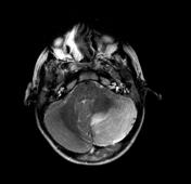
Image 2 MRI (T2) ( create )

Image 3 MRI (FLAIR) ( create )


 Unable to process the form. Check for errors and try again.