
Atrial septal defect
Updates to Article Attributes
Atrial septal defects (ASD) are the second most common congenital heart defect after VSD and the most common to become symptomatic in adulthood.
They are characterised by an abnormal opening in the atrial septum allowing communication between the right and left atria. Due to the low pressures of the atria the lesion is typically asymptomatic until adulthood despite 2-4 times the normal pulmonary blood flow. Gradual(high output) congestive cardiac failure eventually develops, usually becoming symptomatic by the age of 30.
Epidemiology
ASD accounts for ~10% of congenital heart disease 7. There may be greater female predilection.
Clinical presentation
Most patients are asymptomatic but as cardiac failure develops they may present with shortness of breath, palpitations and/or weakness 7.
Pathology
Classification
There are four major types of ASD4, distinguished according to their location within the septum:
-
secundum ASD
- 60-90% of all ASDs
- usually an isolated abnormality
-
primum ASD
- 5-20%
- associated with cleft anterior mitral valve leaflet (partial atrioventricular septal defect)
-
sinus venosus
- 5%
- associated with anomalous right pulmonary venous return to the superior vena cava or right atrium
-
coronary sinus type ASD ("unroofed coronary sinus")
- <1%
- see: unroofed coronary sinus
Associations
ASDs usually tend to be isolated anomalies, associations include:
- Down syndrome (ostium primum defect)
- Holt Oram syndrome
- Ellis van Creveld syndrome
- mitral valve prolapse
- Lutembacher syndrome
- anomalous pulmonary venous return (especially with sinus venosus defects)
Apatent foramen ovale (PFO) is a form of atrial septal defect.
Radiographic features
Plain film (CXR)
- can be normal in
earlyearly stages +/- when the ASD is small - signs of increased pulmonary flow (shunt vascularity)
- enlarged pulmonary vessels
- upper zone vascular prominence
- vessels visible to the periphery of the film
- eventual signs of pulmonary arterial hypertension
- chamber enlargement
- right atrium
- right ventricle
- note: left atrium is normal in size
- note: aortic arch is small to normal
Complications
In approximately 10% of patients pulmonary hypertension develops. In this situation flow through the shunt eventually reverses and becomes right to left. The patient then becomes cyanotic. This is known as theEisenmenger syndrome.
Other complications include:
- paradoxical emboli
- cardiac conduction defects, e.g. atrial fibrillation, flutter.
Treatment and prognosis
ASDs do not cause any impairment in cardiac function in utero and even most neonates are asymptomatic. Either a surgical closure or a percutaneous closure with an Amplatzer closure device can often performed. But careful evaluation has to be made to ensure lack of development of elevated right heart pressures or a right to left shunt prior to any intervention.
-<p><strong>Atrial septal defects (ASD)</strong> are the second most common <a href="/articles/congenital-heart-disease">congenital heart defect</a> after <a href="/articles/ventricular-septal-defect-vsd">VSD</a> and the most common to become symptomatic in adulthood.</p><p>They are characterised by an abnormal opening in the <a href="/articles/atrial-septum">atrial septum</a> allowing communication between the right and left atria. Due to the low pressures of the atria the lesion is typically asymptomatic until adulthood despite 2-4 times the normal pulmonary blood flow. Gradual (high output) congestive cardiac failure eventually develops, usually becoming symptomatic by the age of 30.</p><h4>Epidemiology</h4><p>ASD accounts for ~10% of congenital heart disease <sup>7</sup>. There may be greater female predilection.</p><h4>Clinical presentation</h4><p>Most patients are asymptomatic but as cardiac failure develops they may present with shortness of breath, palpitations and/or weakness <sup>7</sup>. </p><h4>Pathology</h4><h5><strong>Classification</strong></h5><p>There are four major types of ASD <sup>4</sup>, distinguished according to their location within the septum:</p><ul>- +<p><strong>Atrial septal defects (ASD)</strong> are the second most common <a href="/articles/congenital-heart-disease">congenital heart defect</a> after <a href="/articles/ventricular-septal-defect-vsd">VSD</a> and the most common to become symptomatic in adulthood.</p><p>They are characterised by an abnormal opening in the <a href="/articles/atrial-septum">atrial septum</a> allowing communication between the right and left atria. Due to the low pressures of the atria the lesion is typically asymptomatic until adulthood despite 2-4 times the normal pulmonary blood flow. Gradual (high output) congestive cardiac failure eventually develops, usually becoming symptomatic by the age of 30.</p><h4>Epidemiology</h4><p>ASD accounts for ~10% of congenital heart disease <sup>7</sup>. There may be greater female predilection.</p><h4>Clinical presentation</h4><p>Most patients are asymptomatic but as cardiac failure develops they may present with shortness of breath, palpitations and/or weakness <sup>7</sup>. </p><h4>Pathology</h4><h5><strong>Classification</strong></h5><p>There are four major types of ASD <sup>4</sup>, distinguished according to their location within the septum:</p><ul>
-<strong>sinus venosus </strong><ul>- +<strong>sinus venosus </strong><ul>
-<li>associated with anomalous right pulmonary venous return to the <a href="/articles/superior-vena-cava">superior vena cava</a> or <a href="/articles/right-atrium">right atrium </a>- +<li>associated with anomalous right pulmonary venous return to the <a href="/articles/superior-vena-cava">superior vena cava</a> or <a href="/articles/right-atrium">right atrium </a>
-<li>see: <a title="unroofed coronary sinus" href="/articles/unroofed-coronary-sinus">unroofed coronary sinus</a>- +<li>see: <a href="/articles/unroofed-coronary-sinus">unroofed coronary sinus</a>
-</ul><p>A <a href="/articles/patent-foramen-ovale">patent foramen ovale</a> (PFO) is a form of atrial septal defect.</p><h4>Radiographic features</h4><h5>Plain film (CXR)</h5><ul>-<li>can be normal in early stages +/- when the ASD is small</li>- +</ul><p>A <a href="/articles/patent-foramen-ovale">patent foramen ovale</a> (PFO) is a form of atrial septal defect.</p><h4>Radiographic features</h4><h5>Plain film (CXR)</h5><ul>
- +<li>can be normal in early stages +/- when the ASD is small</li>
-</ul><h4>Complications</h4><p>In approximately 10% of patients pulmonary hypertension develops. In this situation flow through the shunt eventually reverses and becomes right to left. The patient then becomes cyanotic. This is known as the <a href="/articles/eisenmenger-syndrome">Eisenmenger syndrome</a>.</p><p>Other complications include:</p><ul>- +</ul><h4>Complications</h4><p>In approximately 10% of patients pulmonary hypertension develops. In this situation flow through the shunt eventually reverses and becomes right to left. The patient then becomes cyanotic. This is known as the <a href="/articles/eisenmenger-syndrome">Eisenmenger syndrome</a>.</p><p>Other complications include:</p><ul>
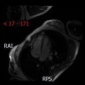
Image 13 MRI (FIESTA) ( create )


 Unable to process the form. Check for errors and try again.