
Cecal volvulus
Updates to Article Attributes
Caecal volvulus describes torsion of the caecum around its own mesentery which often results in obstruction. If unrecognised can result in bowel perforation and faecal peritonitis.
Epidemiology
Caecal volvulus accounts for 11% of all intestinal volvulus and generally occurs in a somewhat younger patients than with a sigmoid volvulus, most being 30-60 years old. There are two predisposing factors that are important for the development of a caecal volvulus:
- There must be developmental failure of peritoneal fixation, allowing the proximal colon to be free and mobile (only occurs in 11-25% of the population).
- Restriction of the bowel at a fixed point within the abdomen that serves as a fulcrum for rotation, e.g. adhesion, abdominal mass, scarring from calcified lymph nodes.
Medical history of these patients may include prior abdominal surgery, presence of a pelvic mass, violent coughing, atonia of the colon, extreme exertion, unpressurized air travel or third-trimester pregnancy.
Clinical presentation
Caecal volvulus presents with clinical features of proximal large bowel obstruction. This is usually with colicky abdominal pain, vomiting and abdominal distension.
Pathology
In approximately half of the patients, the caecum twists in the axial plane, rotating clockwise or counterclockwise around its long axis and appearing in the right lower quadrant.
The other half of patients have what is known as the loop type of cecal volvulus, in which the caecum both twists and inverts, occupying the left upper quadrant of the abdomen. The terminal ileum is usually twisted along with the cecum. Visualization of a gas-filled appendix confirms the diagnosis.
There is a variant of caecal volvulus termed a "caecal bascule" that occurs when the caecum folds anteriorly without any torsion. A caecal bascule is often seen as a dilated loop in the mid-abdomen and although there is an association with prior surgery, adhesions and bands they are not essential for a volvulus to occur. In 10% of the population there is deficient peritoneal fixation of the caecum and ascending colon allowing abnormal mobility. Depending on the length of the mobile segment of right colon a variety of obstructive bowel patterns may result.
Radiographic features
Two types of caecal volvulus are described although, in practice, the distinction is not clinically relevant:
- axial torsion - described below
- caecal bascule
Plain film - abdominal radiograph
Marked distension of a loop of large bowel with its long axis extending from the right lower quadrant to the epigastrium or left upper quadrant. Depending on the initial bowel position and the length of mobile right colon, the distended caecum may be seen anywhere in the abdomen.
Despite the varying positions of the distended cecum, the plain radiographic features of a caecal volvulus are characteristic, and the caput caecum can typically be identified. The colonic haustral pattern is generally maintained in contradistinction to a sigmoid volvulus although some effacement may be present if ischemia develops.
When shorter segments of the colon and cecum are involved, the distended caecum may be found in the normal location. In most patients, obstruction is almost complete and the distal colon is usually empty and the small bowel frequently distended.
Fluoroscopy
Contrast enema
A single-contrast barium or a water soluble contrast enema examination is generally adequate for the evaluation of cecal volvulus. A double-contrast barium enema study does not confer any significant advantage, because no fine detail is necessary to make the diagnosis. The administration of glucagon is often necessary because patients may have considerable colonic spasm and find it difficult to retain the contrast agent.
The barium enema study shows a nondilated distal colon to the point of twist. If the obstruction is not complete, some barium may trickle past the site of obstruction, and the twist may be visualized in more detail. If the twist occurs along the transverse axis, the obstruction appears relatively smooth, and no spiral twist is usually seen. In a cecal bascule, a rounded termination of the barium column may be seen. This, when seen near a distended gas-filled viscus, should alert the radiologist to the diagnosis of a volvulus.
As little barium as possible should be allowed to flow proximal to the site of obstruction because flooding the bowel proximal to the obstruction site might precipitate a complete obstruction. When the barium enema is administered, overdistension should also be avoided because this can lead to perforation. An attempt should always be made to reduce the volvulus. This reduction may be achieved during colonic filling by barium, but reduction occasionally occurs during barium evacuation. With an intermittent volvulus, the barium enema results may be normal, but a postevacuation radiograph may reveal the twist.
CT
On axial CT images, cecal volvulus is suggested by the extreme dilatation of the cecum.
When seen on conventional radiographs or tomograms, the cecal volvulus is seen as a rounded focal collection of air-distended bowel with haustral creases in the left upper quadrant.
The two limbs of the looped obstruction gradually taper and converge at the site of the torsion, resulting in the appearance of a bird's beak. This bird beak sign, originally applied to the appearance of sigmoid volvulus on conventional radiographs, can also be appreciated on axial CT images of the loop-type cecal volvulus.
A specific CT sign for volvulus is the whirl sign, which has been described in volvulus of the midgut, cecum, and sigmoid colon 3. The whirl is composed of spiraled loops of collapsed cecum and sigmoid colon. Low-attenuating fatty mesentery with enhancing engorged vessels radiate from the twisted bowel. In the central eye of the whirl, a soft-tissue density pinpoints the source of the twist. The degree of cecal rotation can even be predicted by the tightness of the whirl.
Treatment and prognosis
Colonoscopic decompression may be appropriate if patient unfit for surgery. However, laparotomy is normally required. Where there is colonic ischaemia, a right hemicolectomy is performed; in some cases, primary anastomosis is not possible and stoma formation at both ends is the safest option.
If the caecum is viable and the volvulus reduces, there are a number of options:
- reduction alone - but this is associated with the highest risk of recurrence
- right hemicolectomy
- caecostomy
- caecopexy
Differential diagnosis
The clinical differential diagnosis is usually between:
- bowel obstruction
- appendicitis
- inflammatory bowel disease
- irritable bowel syndrome
- peptic ulcer disease
The imaging differential is slightly more specific:
- sigmoid volvulus: a sigmoid volvulus arises from the pelvis and is usually ahaustral
See also
-<p><strong>Caecal volvulus</strong> describes torsion of the <a href="/articles/caecum">caecum</a> around its own <a href="/articles/mesentery">mesentery</a> which often results in <a href="/articles/large-bowel-obstruction">obstruction</a>. If unrecognised can result in <a href="/articles/bowel-perforation">bowel perforation</a> and <a href="/articles/faecal-peritonitis">faecal peritonitis</a>. </p><h4>Epidemiology</h4><p>Caecal volvulus accounts for 11% of all intestinal volvulus and generally occurs in a somewhat younger patients than with a <a href="/articles/sigmoid-volvulus">sigmoid volvulus</a>, most being 30-60 years old. There are two predisposing factors that are important for the development of a caecal volvulus:</p><ol>- +<p><strong>Caecal volvulus</strong> describes torsion of the <a href="/articles/caecum">caecum</a> around its own <a href="/articles/mesentery">mesentery</a> which often results in <a href="/articles/large-bowel-obstruction">obstruction</a>. If unrecognised can result in <a href="/articles/bowel-perforation-basic">bowel perforation</a> and <a href="/articles/faecal-peritonitis">faecal peritonitis</a>.</p><h4>Epidemiology</h4><p>Caecal volvulus accounts for 11% of all intestinal volvulus and generally occurs in a somewhat younger patients than with a <a href="/articles/sigmoid-volvulus">sigmoid volvulus</a>, most being 30-60 years old. There are two predisposing factors that are important for the development of a caecal volvulus:</p><ol>
-</ul><h5>Plain film - abdominal radiograph</h5><p>Marked distension of a loop of large bowel with its long axis extending from the right lower quadrant to the epigastrium or left upper quadrant. Depending on the initial bowel position and the length of <a href="/articles/mobile-right-colon">mobile right colon</a>, the distended caecum may be seen anywhere in the abdomen. </p><p>Despite the varying positions of the distended cecum, the plain radiographic features of a caecal volvulus are characteristic, and the <a href="/articles/caput-caecum">caput caecum</a> can typically be identified. The <a href="/articles/colonic-haustral-pattern">colonic haustral pattern</a> is generally maintained in contradistinction to a <a href="/articles/sigmoid-volvulus">sigmoid volvulus</a> although some effacement may be present if ischemia develops. </p><p>When shorter segments of the colon and cecum are involved, the distended caecum may be found in the normal location. In most patients, obstruction is almost complete and the distal colon is usually empty and the small bowel frequently distended. </p><h5>Fluoroscopy</h5><h6>Contrast enema</h6><p>A single-contrast barium or a water soluble contrast enema examination is generally adequate for the evaluation of cecal volvulus. A double-contrast barium enema study does not confer any significant advantage, because no fine detail is necessary to make the diagnosis. The administration of glucagon is often necessary because patients may have considerable colonic spasm and find it difficult to retain the contrast agent.</p><p>The barium enema study shows a nondilated distal colon to the point of twist. If the obstruction is not complete, some barium may trickle past the site of obstruction, and the twist may be visualized in more detail. If the twist occurs along the transverse axis, the obstruction appears relatively smooth, and no spiral twist is usually seen. In a cecal bascule, a rounded termination of the barium column may be seen. This, when seen near a distended gas-filled viscus, should alert the radiologist to the diagnosis of a volvulus.</p><p>As little barium as possible should be allowed to flow proximal to the site of obstruction because flooding the bowel proximal to the obstruction site might precipitate a complete obstruction. When the barium enema is administered, overdistension should also be avoided because this can lead to perforation. An attempt should always be made to reduce the volvulus. This reduction may be achieved during colonic filling by barium, but reduction occasionally occurs during barium evacuation. With an intermittent volvulus, the barium enema results may be normal, but a postevacuation radiograph may reveal the twist.</p><h5>CT</h5><p>On axial CT images, cecal volvulus is suggested by the extreme dilatation of the cecum.</p><p>When seen on conventional radiographs or tomograms, the cecal volvulus is seen as a rounded focal collection of air-distended bowel with haustral creases in the left upper quadrant.</p><p>The two limbs of the looped obstruction gradually taper and converge at the site of the torsion, resulting in the appearance of a bird's beak. This <a href="/articles/bird-beak-sign">bird beak sign</a>, originally applied to the appearance of sigmoid volvulus on conventional radiographs, can also be appreciated on axial CT images of the loop-type cecal volvulus.</p><p>A specific CT sign for volvulus is the <a href="/articles/whirlpool-sign">whirl sign</a>, which has been described in volvulus of the midgut, cecum, and sigmoid colon <sup>3</sup>. The whirl is composed of spiraled loops of collapsed cecum and sigmoid colon. Low-attenuating fatty mesentery with enhancing engorged vessels radiate from the twisted bowel. In the central eye of the whirl, a soft-tissue density pinpoints the source of the twist. The degree of cecal rotation can even be predicted by the tightness of the whirl. </p><h4>Treatment and prognosis</h4><p>Colonoscopic decompression may be appropriate if patient unfit for surgery. However, laparotomy is normally required. Where there is colonic ischaemia, a <a href="/articles/right-hemicolectomy">right hemicolectomy</a> is performed; in some cases, <a href="/articles/primary-anastomosis">primary anastomosis</a> is not possible and stoma formation at both ends is the safest option. </p><p>If the caecum is viable and the volvulus reduces, there are a number of options:</p><ul>- +</ul><h5>Plain film - abdominal radiograph</h5><p>Marked distension of a loop of large bowel with its long axis extending from the right lower quadrant to the epigastrium or left upper quadrant. Depending on the initial bowel position and the length of <a href="/articles/mobile-right-colon">mobile right colon</a>, the distended caecum may be seen anywhere in the abdomen.</p><p>Despite the varying positions of the distended cecum, the plain radiographic features of a caecal volvulus are characteristic, and the <a href="/articles/caput-caecum">caput caecum</a> can typically be identified. The <a href="/articles/colonic-haustral-pattern">colonic haustral pattern</a> is generally maintained in contradistinction to a <a href="/articles/sigmoid-volvulus">sigmoid volvulus</a> although some effacement may be present if ischemia develops.</p><p>When shorter segments of the colon and cecum are involved, the distended caecum may be found in the normal location. In most patients, obstruction is almost complete and the distal colon is usually empty and the small bowel frequently distended.</p><h5>Fluoroscopy</h5><h6>Contrast enema</h6><p>A single-contrast barium or a water soluble contrast enema examination is generally adequate for the evaluation of cecal volvulus. A double-contrast barium enema study does not confer any significant advantage, because no fine detail is necessary to make the diagnosis. The administration of glucagon is often necessary because patients may have considerable colonic spasm and find it difficult to retain the contrast agent.</p><p>The barium enema study shows a nondilated distal colon to the point of twist. If the obstruction is not complete, some barium may trickle past the site of obstruction, and the twist may be visualized in more detail. If the twist occurs along the transverse axis, the obstruction appears relatively smooth, and no spiral twist is usually seen. In a cecal bascule, a rounded termination of the barium column may be seen. This, when seen near a distended gas-filled viscus, should alert the radiologist to the diagnosis of a volvulus.</p><p>As little barium as possible should be allowed to flow proximal to the site of obstruction because flooding the bowel proximal to the obstruction site might precipitate a complete obstruction. When the barium enema is administered, overdistension should also be avoided because this can lead to perforation. An attempt should always be made to reduce the volvulus. This reduction may be achieved during colonic filling by barium, but reduction occasionally occurs during barium evacuation. With an intermittent volvulus, the barium enema results may be normal, but a postevacuation radiograph may reveal the twist.</p><h5>CT</h5><p>On axial CT images, cecal volvulus is suggested by the extreme dilatation of the cecum.</p><p>When seen on conventional radiographs or tomograms, the cecal volvulus is seen as a rounded focal collection of air-distended bowel with haustral creases in the left upper quadrant.</p><p>The two limbs of the looped obstruction gradually taper and converge at the site of the torsion, resulting in the appearance of a bird's beak. This <a href="/articles/bird-beak-sign">bird beak sign</a>, originally applied to the appearance of sigmoid volvulus on conventional radiographs, can also be appreciated on axial CT images of the loop-type cecal volvulus.</p><p>A specific CT sign for volvulus is the <a href="/articles/whirlpool-sign">whirl sign</a>, which has been described in volvulus of the midgut, cecum, and sigmoid colon <sup>3</sup>. The whirl is composed of spiraled loops of collapsed cecum and sigmoid colon. Low-attenuating fatty mesentery with enhancing engorged vessels radiate from the twisted bowel. In the central eye of the whirl, a soft-tissue density pinpoints the source of the twist. The degree of cecal rotation can even be predicted by the tightness of the whirl. </p><h4>Treatment and prognosis</h4><p>Colonoscopic decompression may be appropriate if patient unfit for surgery. However, laparotomy is normally required. Where there is colonic ischaemia, a <a href="/articles/right-hemicolectomy">right hemicolectomy</a> is performed; in some cases, <a href="/articles/primary-anastomosis">primary anastomosis</a> is not possible and stoma formation at both ends is the safest option. </p><p>If the caecum is viable and the volvulus reduces, there are a number of options:</p><ul>
Image ( update )
Image ( update )
Image 1 Photo (Operative photo.) ( update )

Image 2 X-ray (Frontal) ( update )

Image 3 CT (Scanogram) ( update )

Image 4 CT (Scout view) ( update )

Image 5 CT (C+ portal venous phase) ( update )
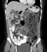
Image 6 X-ray (Frontal) ( update )

Image 7 X-ray (Frontal) ( create )


 Unable to process the form. Check for errors and try again.