
Chest (AP erect view)
Updates to Article Attributes
The erect anteroposterior chest view is the alternative to the PA view when the patient is generally too unwell to tolerate standing or leaving the bed 1. The AP view examines the lungs, bony thoracic cavity mediastinum and great vessels. This particular chest X-ray is often used frequently to aid diagnosis of acute and chronic conditions in intensive care units and wards. The AP view is of lesser quality than the PA view for a number of reasons, yet sometimes it is the only imaging available to that patient.
It is important to note that the AP projection will produce a magnified mediastinal shadow due to the increased distance of the heart from the image receptor and beam divergence ( see figure 3 AP supine and figure 4 PA projection of the same patient) .
Patient position
patient is upright as possible with their back against the image receptor
the chin is raised as to be out of the image field
if possible the hands are placed by the patient's side
shoulders are depressed to move the clavicles below the lung apices
Technical factors
anteroposterior projection
suspended inspiration
-
centring point
the level of the 7th thoracic vertebra, approximately 7 cm below the jugular notch of the sternum
The central ray is angled to be perpendicular to the long axis of the patient's sternum generally resulting in an caudal angle
-
collimation
superiorly 5 cm above the shoulder joint to allow proper visualisation of the upper airways
inferior to the inferior border of the 12th rib
lateral to the level of the acromioclavicular joints
-
orientation
portrait or landscape
-
detector size
35 cm x 43 cm or 43 cm x 35 cm
-
exposure
100-110 kVp
4-8 mAs
-
SID
180 cm
-
grid
yes (this may be departmentally dependent)
Image technical evaluation
The entire lung fields should be visible from the apices down to the lateral costophrenic angles.
three posterior ribs should be seen above the superior aspect of the clavicle
the chin should not be superimposing any structures
sternoclavicular joints are equal distant apart
the clavicle are in the same horizontal plane
a minimum of eight posterior ribs are visualised above the diaphragm
the ribs and thoracic cage are seen only faintly over the heart
clear vascular markings of the lungs should be visible
Practical points
This projection can be very challenging in emergency situations, clear communication is the key to ensure your patient gets the best image possible under the situation at hand.
The AP view, although a supplementary projection for the PA comes with a wide range of technically challenging factors and is hence inferior.
By and large AP erect patients are quite unwell and may not have the ability to aid the radiographer in positioning. The projection can be done in the wards using a mobile machine or in the general rooms using a portable image receptor.
When sitting the patient up for the x ray it is important to explain not only to the patient but the staff around; that you will be sitting that patient quite upright for their radiograph, often this will require aid.
There will be occasions where the patient cannot hold themselves upright, in these situations it is not uncommon to wedge pillows in between the patient and the bedside to prop them up or ultimately resort to a supine chest projection.
As a general rule, there will be a rough 10-15° caudal angle although in heavily kyphotic patients no angle may be required.
If the clavicles are projected overly inferior less angle is required, if the clavicles are projected above the apices more angle should be applied to obtain a diagnostic image.
The phase of respiration has a profound effect on the appearance of several structures on the chest radiograph. A poor-inspiratory AP radiograph can mimic pathology. Structures that can appear different on expiration include:
heart size
mediastinal contours and width
lung inflation
diaphragm contours
Rotation of a chest radiograph can mimic common pathology processes and make it hard to produce a pertinent diagnosis.
The AP view is used to investigate a plethora of conditions and it is the radiographer's responsibility to ensure high quality diagnostic images are achieved consistently.
The sternoclavicular joints are a sound indicator for positional rotation, if one sternoclavicular joint is notably wider than the other, that respected side needs to be rotated away from the image receptor to correct rotation.
Patients with long standing history of emphysema or COPD will have abnormally long lungs compared to the general population, remember this when collimating superior to inferior.
Side marker placement is imperative, patients can have congenital conditions that mimic a mirrored image 2.
Remember to explain to your patient what you are about to do, that is ask them to take a breath in and hold it. Many times this gives the patient time to prepare and results in a better breath hold and therefore a higher quality radiograph.
Always remember to tell your patient to breathe again!
-<p>The <strong>erect anteroposterior chest view </strong>is the alternative to the PA view when the patient is generally too unwell to tolerate standing or leaving the bed <sup>1</sup>. The AP view examines the <a href="/articles/lung">lungs</a>, bony thoracic cavity<a href="/articles/normal-contours-of-the-cardiomediastinum-on-chest-radiography"> mediastinum</a> and <a href="/articles/great-vessel-space-1">great vessels</a>. This particular chest X-ray is often used frequently to aid diagnosis of acute and chronic conditions in intensive care units and wards. The AP view is of lesser quality than the PA view for a number of reasons, yet sometimes it is the only imaging available to that patient.</p><p>It is important to note that the AP projection will produce a magnified mediastinal shadow due to the increased distance of the heart from the image receptor and beam divergence ( see figure 3 <em>AP supine</em> and figure 4 <em>PA projection</em> of the same patient) .</p><h4><strong>Patient position</strong></h4><ul>- +<p>The <strong>erect anteroposterior chest view </strong>is the alternative to the PA view when the patient is generally too unwell to tolerate standing or leaving the bed <sup>1</sup>. The AP view examines the <a href="/articles/lung">lungs</a>, bony thoracic cavity<a href="/articles/normal-contours-of-the-cardiomediastinum-on-chest-radiography"> mediastinum</a> and <a href="/articles/great-vessel-space-1">great vessels</a>. This particular chest X-ray is often used frequently to aid diagnosis of acute and chronic conditions in intensive care units and wards. The AP view is of lesser quality than the PA view for a number of reasons, yet sometimes it is the only imaging available to that patient.</p><p>It is important to note that the AP projection will produce a magnified mediastinal shadow due to the increased distance of the heart from the image receptor and beam divergence ( see figure 3 <em>AP supine</em> and figure 4 <em>PA projection</em> of the same patient) .</p><h4>Patient position</h4><ul>
-</ul><h4><strong>Technical factors</strong></h4><ul>- +</ul><h4>Technical factors</h4><ul>
-</ul><h4><strong>Image technical evaluation</strong></h4><p>The entire lung fields should be visible from the<a href="/articles/apical-zone"> apices</a> down to the lateral costophrenic angles.</p><ul>- +</ul><h4>Image technical evaluation</h4><p>The entire lung fields should be visible from the<a href="/articles/apical-zone"> apices</a> down to the lateral costophrenic angles.</p><ul>
-</ul><h4><strong>Practical points</strong></h4><p>This projection can be very challenging in emergency situations, clear communication is the key to ensure your patient gets the best image possible under the situation at hand.</p><p>The AP view, although a supplementary projection for the PA comes with a wide range of technically challenging factors and is hence inferior.</p><p>By and large AP erect patients are quite unwell and may not have the ability to aid the radiographer in positioning. The projection can be done in the wards using a mobile machine or in the general rooms using a portable image receptor.</p><p>When sitting the patient up for the x ray it is important to explain not only to the patient but the staff around; that you will be sitting that patient quite upright for their radiograph, often this will require aid.</p><p>There will be occasions where the patient cannot hold themselves upright, in these situations it is not uncommon to wedge pillows in between the patient and the bedside to prop them up or ultimately resort to a supine chest projection.</p><p>As a general rule, there will be a rough 10-15° caudal angle although in heavily <a title="kyphotic deformity" href="/articles/kyphosis">kyphotic</a> patients no angle may be required.</p><p>If the clavicles are projected overly inferior less angle is required, if the clavicles are projected above the apices more angle should be applied to obtain a diagnostic image.</p><p>The <strong>phase of respiration</strong> has a profound effect on the appearance of several structures on the chest radiograph. A poor-inspiratory AP radiograph can mimic pathology. Structures that can appear different on expiration include:</p><ul>- +</ul><h4>Practical points</h4><p>This projection can be very challenging in emergency situations, clear communication is the key to ensure your patient gets the best image possible under the situation at hand.</p><p>The AP view, although a supplementary projection for the PA comes with a wide range of technically challenging factors and is hence inferior.</p><p>By and large AP erect patients are quite unwell and may not have the ability to aid the radiographer in positioning. The projection can be done in the wards using a mobile machine or in the general rooms using a portable image receptor.</p><p>When sitting the patient up for the x ray it is important to explain not only to the patient but the staff around; that you will be sitting that patient quite upright for their radiograph, often this will require aid.</p><p>There will be occasions where the patient cannot hold themselves upright, in these situations it is not uncommon to wedge pillows in between the patient and the bedside to prop them up or ultimately resort to a supine chest projection.</p><p>As a general rule, there will be a rough 10-15° caudal angle although in heavily <a href="/articles/kyphosis">kyphotic</a> patients no angle may be required.</p><p>If the clavicles are projected overly inferior less angle is required, if the clavicles are projected above the apices more angle should be applied to obtain a diagnostic image.</p><p>The <strong>phase of respiration</strong> has a profound effect on the appearance of several structures on the chest radiograph. A poor-inspiratory AP radiograph can mimic pathology. Structures that can appear different on expiration include:</p><ul>
References changed:
- 1. Jr RBJ, FACR BJMMDP, Osborn AG et-al. Diagnostic Imaging: Emergency: Published by Amirsys. Lippincott Williams & Wilkins. ISBN:1931884765. <a href="http://books.google.com/books?vid=ISBN1931884765">Read it at Google Books</a> - <a href="http://www.amazon.com/gp/product/1931884765">Find it at Amazon</a><span class="auto"></span>
- 2. Keats TE, Anderson MW. Atlas of Normal Roentgen Variants That May Simulate Disease. Saunders. (2012) ISBN:0323073557. <a href="http://books.google.com/books?vid=ISBN0323073557">Read it at Google Books</a> - <a href="http://www.amazon.com/gp/product/0323073557">Find it at Amazon</a><span class="ref_v3"></span>
Sections changed:
- Radiography
Systems changed:
- Chest
Image 1 X-ray (Frontal) ( create )
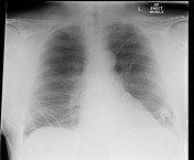
Image 2 X-ray (Frontal) ( create )

Image 3 X-ray (Frontal) ( create )

Image 4 X-ray (Frontal) ( create )


 Unable to process the form. Check for errors and try again.