
Chest (PA view)
Updates to Article Attributes
Chest: PA view is the most common radiological investigation in the emergency department 1. The PA view examines the lungs, bony thoracic cavity, mediastinum and great vessels. The chest X-ray is used frequently to aid diagnosis of acute and chronic conditions.
Patient position
- patient is erect facing the upright image receptor, the superior aspect of the receptor is 5 cm above the shoulder joints
- the chin is raised as to be out of the image field
- shoulders are rotated anteriorly to allow the scapulae to move laterally off the lung fields, this can be achieved by either:
- hands placed on the posterior aspect of the hips, elbows partially flexed rolling anterior or
- hands are placed around the image receptor in a hugging motion with focus on lateral movement of the scapulae
- shoulders are depressed to move the clavicles below the lung apices
Technical factors
- posterioranterior projection
- suspended inspiration
-
centring point
- the level of the 7th thoracic vertebra, approximately the inferior angle of the scapulae
-
collimation
- superiorly 5 cm above the shoulder joint to allow proper visualisation of the upper airways
- inferior to the inferior border of the 12th rib
- lateral to the level of the acromioclavicular joints
-
orientation
- portrait or landscape
-
detector size
- 35 cm x 43 cm or 43 cm x 35 cm
-
exposure
- 100-110 kVp
- 4-8 mAs
-
SID
- 180 cm
-
grid
- yes
Image technical evaluation
The entire lung fields should be visible from the apices down to the lateral costophrenic angles.
The chin should not be superimposing any structures.
Minimal to no superimposition of the scapulae boards on the lung fields.
Sternoclavicular joints are equal distant apart.
The clavicle are in the same horizontal plane.
A minimum of ten posterior ribs are visualised above the diaphragm.
The ribs and thoracic cage are seen only faintly over the heart.
Clear vascular markings of the lungs should be visible.
Practical points
Rotation of a PA chest X-ray can mimic common disease processes and make it hard to produce a pertinent diagnosis.
The PA view is used to investigate a plethora of conditions and it is the radiographer's responsibility to ensure high quality diagnostic images are achieved consistently.
The medial aspect of the clavicles are a good indicator of rotation, if the medial clavicle is projected over the trachea the opposing side must be turned anterior. For example, if the medial end of the left clavicle is veering to the right of the patient projecting over the trachea; rotating the patient anteriorly to the right will correct this.
Patients with longstanding history of emphysema or COPD will have abnormally long lungs compared to the general population, remember this when collimating superior to inferior.
Side marker placement is imperative, patients can have congenital conditions that mimic a mirrored image.
Remember to explain to your patient what you are about to do, that is ask them to take a breath in and hold it. Many times this gives the patient time to prepare and results in a better breath hold and therefore a higher quality radiograph.
Always remember to tell your patient to breathbreathe again!
-<p> </p><p><strong>Chest: PA view </strong>is the most common radiological investigation in the emergency department <sup>1</sup>. The PA view examines the <a title="Lungs" href="/articles/lung">lungs</a>, bony thoracic cavity, <a title="Normal contours of the cardiomediastinum on chest radiography" href="/articles/normal-contours-of-the-cardiomediastinum-on-chest-radiography">mediastinum </a>and <a title="Great vessel space" href="/articles/great-vessel-space-1">great vessels</a>. The chest X-ray is used frequently to aid diagnosis of acute and chronic conditions. </p><h4>Patient position</h4><ul>- +<p> </p><p><strong>Chest: PA view </strong>is the most common radiological investigation in the emergency department <sup>1</sup>. The PA view examines the <a href="/articles/lung">lungs</a>, bony thoracic cavity, <a href="/articles/normal-contours-of-the-cardiomediastinum-on-chest-radiography">mediastinum </a>and <a href="/articles/great-vessel-space-1">great vessels</a>. The chest X-ray is used frequently to aid diagnosis of acute and chronic conditions. </p><h4>Patient position</h4><ul>
-<strong>centring point</strong><ul><li>the level of the<a title="Typical thoracic vertebrae" href="/articles/typical-thoracic-vertebrae"> 7<sup>th</sup> thoracic vertebra</a>, approximately the inferior angle of the scapulae </li></ul>- +<strong>centring point</strong><ul><li>the level of the<a href="/articles/typical-thoracic-vertebrae"> 7<sup>th</sup> thoracic vertebra</a>, approximately the inferior angle of the scapulae </li></ul>
-<li>lateral to the level of the <a title="Acromioclavicular joint configuration" href="/articles/acromioclavicular-joint-configuration">acromioclavicular joints</a>- +<li>lateral to the level of the <a href="/articles/acromioclavicular-joint-configuration">acromioclavicular joints</a>
-</ul><h4>Image technical evaluation </h4><p>The entire lung fields should be visible from the <a title="Apices" href="/articles/apical-zone">apices</a> down to the lateral costophrenic angles. </p><p>The chin should not be superimposing any structures.</p><p>Minimal to no superimposition of the scapulae boards on the lung fields.</p><p><a title="Sternoclavicular joint" href="/articles/sternoclavicular-joint">Sternoclavicular</a> joints are equal distant apart. </p><p>The <a title="Clavicle anatomy" href="/articles/clavicle">clavicle</a> are in the same horizontal plane.</p><p>A minimum of ten posterior ribs are visualised above the diaphragm.</p><p>The ribs and thoracic cage are seen only faintly over the heart. </p><p>Clear vascular markings of the lungs should be visible. </p><h4>Practical points</h4><p>Rotation of a PA chest X-ray can mimic common disease processes and make it hard to produce a pertinent diagnosis.</p><p>The PA view is used to investigate a plethora of conditions and it is the radiographer's responsibility to ensure high quality diagnostic images are achieved consistently.</p><p>The medial aspect of the clavicles are a good indicator of rotation, if the medial clavicle is projected over the trachea the opposing side must be turned anterior. For example, if the medial end of the left clavicle is veering to the right of the patient projecting over the trachea; rotating the patient anteriorly to the right will correct this. </p><p>Patients with longstanding history of <a title="Emphysema of the lung" href="/articles/pulmonary-emphysema">emphysema</a> or <a title="COPD (basic)" href="/articles/copd-basic">COPD</a> will have abnormally long lungs compared to the general population, remember this when collimating superior to inferior.</p><p>Remember to explain to your patient what you are about to do, that is ask them to take a breath in and hold it. Many times this gives the patient time to prepare and results in a better breath hold and therefore a higher quality radiograph.</p><p>Always remember to tell your patient to breath again!</p><p> </p>- +</ul><h4>Image technical evaluation </h4><p>The entire lung fields should be visible from the <a href="/articles/apical-zone">apices</a> down to the lateral costophrenic angles. </p><p>The chin should not be superimposing any structures.</p><p>Minimal to no superimposition of the scapulae boards on the lung fields.</p><p><a href="/articles/sternoclavicular-joint">Sternoclavicular</a> joints are equal distant apart. </p><p>The <a href="/articles/clavicle">clavicle</a> are in the same horizontal plane.</p><p>A minimum of ten posterior ribs are visualised above the diaphragm.</p><p>The ribs and thoracic cage are seen only faintly over the heart. </p><p>Clear vascular markings of the lungs should be visible. </p><h4>Practical points</h4><p>Rotation of a PA chest X-ray can mimic common disease processes and make it hard to produce a pertinent diagnosis.</p><p>The PA view is used to investigate a plethora of conditions and it is the radiographer's responsibility to ensure high quality diagnostic images are achieved consistently.</p><p>The medial aspect of the clavicles are a good indicator of rotation, if the medial clavicle is projected over the trachea the opposing side must be turned anterior. For example, if the medial end of the left clavicle is veering to the right of the patient projecting over the trachea; rotating the patient anteriorly to the right will correct this. </p><p>Patients with longstanding history of <a href="/articles/pulmonary-emphysema">emphysema</a> or <a href="/articles/copd-basic">COPD</a> will have abnormally long lungs compared to the general population, remember this when collimating superior to inferior.</p><p>Side marker placement is imperative, patients can have <a href="/articles/situs-inversus">congenital conditions</a> that mimic a mirrored image. </p><p>Remember to explain to your patient what you are about to do, that is ask them to take a breath in and hold it. Many times this gives the patient time to prepare and results in a better breath hold and therefore a higher quality radiograph.</p><p>Always remember to tell your patient to breathe again!</p><p> </p>
References changed:
- 1. Jr RBJ, FACR BJMMDP, Osborn AG et-al. Diagnostic Imaging: Emergency: Published by Amirsys. Lippincott Williams & Wilkins. ISBN:1931884765. <a href="http://books.google.com/books?vid=ISBN1931884765">Read it at Google Books</a> - <a href="http://www.amazon.com/gp/product/1931884765">Find it at Amazon</a><span class="auto"></span>
Sections changed:
- Radiography
Systems changed:
- Chest
Image ( create )
Image ( create )
Image 1 X-ray (Frontal) ( create )
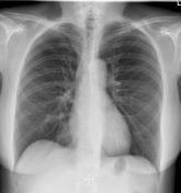
Image 2 X-ray (Frontal insp) ( create )


 Unable to process the form. Check for errors and try again.