
Consolidation
Updates to Article Attributes
Air space opacification is a descriptive term that refers to the filling of the pulmonary tree with material that attenuates x-rays more than the surrounding lung parenchyma. It is one of the many patterns of lung opacification and is equivalent to the pathological diagnosis of pulmonary consolidation.
In radiological studies, it presents as increased attenuation of the lung parenchyma causing obscuration of pulmonary vessels, without significant loss of volume, in the segment(s) affected. Air bronchograms can also be found 1, 2.
Pathology
CausesAetiology
The opacification is caused by fluid or solid material within the airways that causes a difference in the relative attenuation of the lung:
- transudate, e.g. pulmonary oedema secondary to heart failure
- pus, e.g. bacterial pneumonia
- blood, e.g. pulmonary haemorrhage
- cells, e.g. bronchoalveolar carcinoma
- protein, e.g. alveolar proteinosis
- fat, e.g. lipoid pneumonia
- gastric contents, e.g. aspiration pneumonia
- water, e.g. drowning
When considering the likely causes of airspace opacification, it is useful to determine chronicity (by reviewing previous radiographs) and considering laterality.
Additionally, the presence of mediastinal or hilar lymphadenopathy further refines the massive list of differentials:
- acute unilateral air space opacification
- acute bilateral air space opacification
- acute airspace opacification with lymphadenopathy
- chronic unilateral airspace opacification
- chronic bilateral airspace opacification
Patterns of disease
On chest radiography a number of patterns are recognised:
-<p><strong>Air space opacification </strong>is a descriptive term that refers to filling of the pulmonary tree with material that attenuates x-rays more than the surrounding <a href="/articles/lung-parenchyma">lung parenchyma</a>. It is one of the many <a href="/articles/pulmonary-opacification">patterns of lung opacification</a> and is equivalent to the pathological diagnosis of <strong>pulmonary consolidation</strong>.</p><p>In radiological studies, it presents as increased attenuation of the lung parenchyma causing obscuration of pulmonary vessels, without significant loss of volume, in the segment(s) affected. <a href="/articles/air-bronchogram">Air bronchograms</a> can also be found <sup>1, 2</sup>.</p><h4>Pathology</h4><h5>Causes</h5><p>The opacification is caused by fluid or solid material within the airways that causes a difference in the relative attenuation of the lung:</p><ul>- +<p><strong>Air space opacification </strong>is a descriptive term that refers to the filling of the pulmonary tree with material that attenuates x-rays more than the surrounding <a href="/articles/lung-parenchyma">lung parenchyma</a>. It is one of the many <a href="/articles/pulmonary-opacification">patterns of lung opacification</a> and is equivalent to the pathological diagnosis of <strong>pulmonary consolidation</strong>.</p><p>In radiological studies, it presents as increased attenuation of the lung parenchyma causing obscuration of pulmonary vessels, without significant loss of volume, in the segment(s) affected. <a href="/articles/air-bronchogram">Air bronchograms</a> can also be found <sup>1, 2</sup>.</p><h4>Pathology</h4><h5>Aetiology</h5><p>The opacification is caused by fluid or solid material within the airways that causes a difference in the relative attenuation of the lung:</p><ul>
References changed:
- 1. Kuhlman J, Scatarige J, Fishman E, Zerhouni E, Siegelman S. CT Demonstration of High Attenuation Pleural-Parenchymal Lesions Due to Amiodarone Therapy. J Comput Assist Tomogr. 1987;11(1):160-2. <a href="https://doi.org/10.1097/00004728-198701000-00034">doi:10.1097/00004728-198701000-00034</a> - <a href="https://www.ncbi.nlm.nih.gov/pubmed/3805405">Pubmed</a>
- 2. Silva C, Marchiori E, Souza Júnior A, Müller N. Illustrated Brazilian Consensus of Terms and Fundamental Patterns in Chest CT Scans. J Bras Pneumol. 2010;36(1):99-123. <a href="https://doi.org/10.1590/s1806-37132010000100016">doi:10.1590/s1806-37132010000100016</a> - <a href="https://www.ncbi.nlm.nih.gov/pubmed/20209314">Pubmed</a>
- 1. Kuhlman JE, Scatarige JC, Fishman EK et-al. CT demonstration of high attenuation pleural-parenchymal lesions due to amiodarone therapy. J Comput Assist Tomogr. 1987;11 (1): 160-2. <a href="http://www.ncbi.nlm.nih.gov/pubmed/3805405">Pubmed citation</a><span class="ref_v3"></span>
- 2. Silva CI, Marchiori E, Souza JúNior AS et-al. Illustrated Brazilian consensus of terms and fundamental patterns in chest CT scans. J Bras Pneumol.;36 (1): 99-123. <a href="http://www.scielo.br/scielo.php?script=sci_arttext&pid=S1806-37132010000100016&lng=pt&nrm=iso&tlng=pt">J Bras Pneumol (full text)</a> - <a href="http://dx.doi.org/10.1590/S1806-37132010000100016">doi:10.1590/S1806-37132010000100016</a> - <a href="http://www.ncbi.nlm.nih.gov/pubmed/20209314">Pubmed citation</a><span class="ref_v3"></span>
Image 2 CT (lung window) ( update )

Image 3 X-ray (Frontal) ( update )

Image 4 X-ray (Frontal) ( update )

Image 5 CT (lung window) ( update )
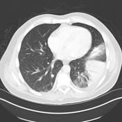
Image 6 X-ray (Frontal) ( update )

Image 7 Annotated image ( update )


 Unable to process the form. Check for errors and try again.