
Diaphragmatic paralysis
Updates to Article Attributes
Diaphragmatic paralysis can be unilateral or bilateral.
Clinical presentation
Clinical features are highly variable according to underlying aetiological factor:
- unilateral paralysis: asymptomatic in most of the patients as the other lung compensates
- may have dyspnoea, headaches, fatigue, insomnia and overall breathing difficulty
- bilateral diaphragmatic palsy can be a medical emergency; they present with severe dyspnoea, even with mild exertion
Pathology
Aetiology
- idiopathic: accounts for ~70% of the cases
-
phrenic nerve palsy, which in turn can occur from
- bronchogenic carcinoma and other neoplasms
- spinal cord pathology
- spinal cord injury
- myelitis
- cardiac surgery7
- Erb's palsy (birth trauma)
- muscular disorders
- iatrogenic causes
- cerebral hypoventilation syndrome (Ondine's curse)
Radiographic features
Plain radiograph
Normally the right dome of the diaphragm is higher in position as compared to the left dome, if the left dome of the diaphragm is elevated (>2 cm) diaphragmatic palsy should be suspected.
Fluoroscopy
Fluoroscopic examination of the diaphragm ("sniff test") is very useful in diagnosing a diaphragmatic paralysis. In normal individuals, both hemidiaphragm will descend with inspiration. In cases of unilateral diaphragmatic paralysis, the affected side demonstrates a paradoxical upward movement.
Ultrasound
An alternative to fluoroscopy in diagnosing this condition, particularly useful in the paediatric population. Real-time ultrasound is ideal for evaluation of spontaneous respiratory diaphragmatic motion (may require temporary disconnection of the ventilator). This can be performed in the axial plane to compare the two hemidiaphragm simultaneously. Additional coronal or sagittal M-mode can help quantify the degree of movement of each individual hemidiaphragm. Diagnostic criteria include paradoxical movement, excursion of less than 4 mm, and a difference >50% between the excursion of one hemidiaphragm compared to the other.
Treatment and prognosis
Patients with unilateral diaphragmatic paralysis do not require treatment. There may be an option for phrenic nerve stimulation in some cases.
Differential diagnosis
On a chest radiograph consider:
- right diaphragmatic eventration
- lobar collapse
- subphrenic abscess
- subdiaphragmatic mass
-<li>cardiac surgery</li>- +<li>cardiac surgery <sup>7</sup>
- +</li>
-</ul><h4>Radiographic features</h4><h5>Plain radiograph</h5><p>Normally the right dome of the diaphragm is higher in position as compared to the left dome, if the left dome of the diaphragm is elevated (>2 cm) diaphragmatic palsy should be suspected.</p><h5>Fluoroscopy</h5><p>Fluoroscopic examination of the diaphragm ("<a href="/articles/sniff-test">sniff test</a>") very useful in diagnosing a diaphragmatic paralysis. In normal individuals, both hemidiaphragm will descend with inspiration. In cases of unilateral diaphragmatic paralysis, the affected side demonstrates a paradoxical upward movement.</p><h5>Ultrasound</h5><p>An alternative to fluoroscopy in diagnosing this condition, particularly useful in the paediatric population. Real-time ultrasound is ideal for evaluation of spontaneous respiratory diaphragmatic motion (may require temporary disconnection of the ventilator). This can be performed in the axial plane to compare the two hemidiaphragm simultaneously. Additional coronal or sagittal M-mode can help quantify the degree of movement of each individual hemidiaphragm. Diagnostic criteria include paradoxical movement, excursion of less than 4 mm, and a difference >50% between the excursion of one hemidiaphragm compared to the other.</p><h4>Treatment and prognosis</h4><p>Patients with unilateral diaphragmatic paralysis do not require treatment. There may be an option for <a href="/articles/phrenic-nerve-stimulation">phrenic nerve stimulation</a> in some cases.</p><h4>Differential diagnosis</h4><p>On a chest radiograph consider:</p><ul>- +</ul><h4>Radiographic features</h4><h5>Plain radiograph</h5><p>Normally the right dome of the diaphragm is higher in position as compared to the left dome, if the left dome of the diaphragm is elevated (>2 cm) diaphragmatic palsy should be suspected.</p><h5>Fluoroscopy</h5><p>Fluoroscopic examination of the diaphragm ("<a href="/articles/sniff-test">sniff test</a>") is very useful in diagnosing a diaphragmatic paralysis. In normal individuals, both hemidiaphragm will descend with inspiration. In cases of unilateral diaphragmatic paralysis, the affected side demonstrates a paradoxical upward movement.</p><h5>Ultrasound</h5><p>An alternative to fluoroscopy in diagnosing this condition, particularly useful in the paediatric population. Real-time ultrasound is ideal for evaluation of spontaneous respiratory diaphragmatic motion (may require temporary disconnection of the ventilator). This can be performed in the axial plane to compare the two hemidiaphragm simultaneously. Additional coronal or sagittal M-mode can help quantify the degree of movement of each individual hemidiaphragm. Diagnostic criteria include paradoxical movement, excursion of less than 4 mm, and a difference >50% between the excursion of one hemidiaphragm compared to the other.</p><h4>Treatment and prognosis</h4><p>Patients with unilateral diaphragmatic paralysis do not require treatment. There may be an option for <a href="/articles/phrenic-nerve-stimulation">phrenic nerve stimulation</a> in some cases.</p><h4>Differential diagnosis</h4><p>On a chest radiograph consider:</p><ul>
References changed:
- 6. Harriet Paltiel. Pediatric Ultrasound, An Issue of Ultrasound Clinics,. (2013) <a href="https://books.google.co.uk/books?vid=ISBN9781455773701">ISBN: 9781455773701</a><span class="ref_v4"></span>
- 7. Talwar S, Agarwala S, Mittal C, Choudhary S, Airan B. Diaphragmatic Palsy After Cardiac Surgical Procedures in Patients with Congenital Heart. Ann Pediatr Card. 2010;3(1):50. <a href="https://doi.org/10.4103/0974-2069.64370">doi:10.4103/0974-2069.64370</a> - <a href="https://www.ncbi.nlm.nih.gov/pubmed/20814476">Pubmed</a>
- 6. Paltiel, Harriet. Pediatric Ultrasound, An Issue of Ultrasound Clinics. Volume 8, Issue 3 of The Clinics: Radiology. Elsevier Health Sciences, 2013
Image 2 X-ray (Frontal) ( update )

Image 4 X-ray (Frontal) ( update )

Image 5 CT (lung window) ( update )

Image 6 X-ray (Frontal) ( update )
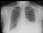
Image 7 CT (lung window) ( create )


 Unable to process the form. Check for errors and try again.