
Diffusion tensor imaging and fiber tractography
Updates to Article Attributes
Diffusion-tensor tensor imaging (DTI) is an MRI technique that uses anisotropic diffusion to estimate the axonal (white matter) organisation of the brain.
FiberFibre tractography (FT) is a 3D reconstruction technique to accessassess neural tracts using data collected by DTIdiffusion tensor imaging.
Diffusion-weighted imaging (DWI) is based on the measurement of thermal Brownian motion of water molecules. Within cerebral white matter, water molecules tend to diffuse more freely along the direction of axonal fascicles rather than across them. Such directional dependence of diffusivity is termed anisotropy. This direction of maximum diffusivity along the white-matter fibres is projected in the final image.
Colour coding Clinical applications
For many, diffusion tensor imaging is synonymous with MR imaging of the CNS. However, its use for the assessment of highly-organised body systems outside the CNS, where anisotropy can facilitate early detection of pathology, has been gaining favour in recent years. Applications include:
-
red for fibres crossing from left to rightassessment of the deformation of white matter by tumours - deviation, infiltration, destruction of white matter -
green for fibres traversing in anteroposterior directiondelineation of the anatomy of immature brains -
blue for fibres going from superior to inferiorpre-surgical planning - Alzheimer disease - detection of early disease
- schizophrenia
- focal cortical dysplasia
- multiple sclerosis - plaque assessment
- early identification of musculoskeletal and peripheral nerve pathology 7-10
Quantitative analysis methods
- ROI (region of interest) based
- voxel-based
- histogram analysis
- tractography/fibre tracking
Physics of DTI
Some terms
-
DTI providesDiffusion tensor imaging provides a quantitative analysis of the magnitude and directionality of water molecules - the word tensor indicates the use of a
3 x 33x3 matrixused herewitheigen valueseigenvalues andeigen vectorseigenvectors as its constituents - the two main parameters derived from DTI data are mean diffusivity (MD)
or in another term, also referred to as apparent diffusion coefficient (ADC), and fractional anisotropy (FA) - FA reflects the directionality of molecular displacement by diffusion and
varyvaries between 0 (isotropic diffusion) and 1 (infinite anisotropic diffusion). FA value of CSF is 0 - MD reflects the average magnitude of molecular displacement by diffusion. The
morehigher the MD value, the moretheisotropicisthe medium - AD - axial diffusivity represents the longest
eigen vectoreigenvector - RD - radial diffusivity represents the average of two shorter
eigen vectorseigenvectors - DTI data acquisition is done by SE-EPI with the application of diffusion gradients in multiple directions (SE-EPI = spin-echo echo-planar imaging, using pairs of 90-180 degree pulses, giving T2-weighting)
Colour coding
In fibre tractography imaging, the convention for colour coding is a follows:
- red: transverse fibres
- green: anteroposterior fibres
- blue: craniocaudal fibres
-<p><strong>Diffusion-tensor imaging (DTI) </strong>is an MRI technique that uses anisotropic diffusion to estimate the axonal (white matter) organisation of the brain. <strong>Fiber tractography (FT)</strong> is a 3D reconstruction technique to access neural tracts using data collected by DTI.</p><p>Within cerebral white matter, water molecules tend to diffuse more freely along the direction of axonal fascicles than across them. Such directional dependence of diffusivity is termed anisotropy.</p><h4>Colour coding </h4><ul>-<li>red for fibres crossing from left to right</li>-<li>green for fibres traversing in anteroposterior direction</li>-<li>blue for fibres going from superior to inferior</li>- +<p><strong>Diffusion tensor imaging (DTI) </strong>is an MRI technique that uses anisotropic diffusion to estimate the axonal (white matter) organisation of the brain.</p><p><strong>Fibre tractography (FT)</strong> is a 3D reconstruction technique to assess neural tracts using data collected by diffusion tensor imaging.</p><p><a href="/articles/diffusion-weighted-imaging-1">Diffusion-weighted imaging</a> (DWI) is based on the measurement of thermal Brownian motion of water molecules. Within cerebral white matter, water molecules tend to diffuse more freely along the direction of axonal fascicles rather than across them. Such directional dependence of diffusivity is termed <a href="/articles/anisotropy">anisotropy</a>. This direction of maximum diffusivity along the white-matter fibres is projected in the final image.</p><h4>Clinical applications</h4><p>For many, diffusion tensor imaging is synonymous with MR imaging of the CNS. However, its use for the assessment of highly-organised body systems outside the CNS, where anisotropy can facilitate early detection of pathology, has been gaining favour in recent years. Applications include:</p><ul>
- +<li>assessment of the deformation of white matter by tumours - deviation, infiltration, destruction of white matter</li>
- +<li>delineation of the anatomy of immature brains</li>
- +<li>pre-surgical planning</li>
- +<li>
- +<a href="/articles/alzheimer-disease-1">Alzheimer disease</a> - detection of early disease</li>
- +<li><a href="/articles/schizophrenia">schizophrenia</a></li>
- +<li><a href="/articles/focal-cortical-dysplasia">focal cortical dysplasia</a></li>
- +<li>
- +<a href="/articles/multiple-sclerosis">multiple sclerosis</a> - plaque assessment</li>
- +<li>early identification of musculoskeletal and peripheral nerve pathology <sup>7-10</sup>
- +</li>
-</ul><h4>Physics of DTI</h4><ul>-<li>DTI provides quantitative analysis of the magnitude and directionality of water molecules</li>-<li>the word tensor indicates a 3 x 3 matrix used here with eigen values and eigen vectors as its constituents</li>-<li>the two main parameters derived from DTI data are mean diffusivity (MD) or in another term apparent diffusion coefficient (ADC) and fractional anisotropy (FA)</li>-<li>FA reflects the directionality of molecular displacement by diffusion and vary between 0 (isotropic diffusion) and 1 (infinite anisotropic diffusion). FA value of CSF is 0</li>-<li>MD reflects the average magnitude of molecular displacement by diffusion. The more the MD value, the more the isotropic is the medium</li>-<li>AD - axial diffusivity represents the longest eigen vector </li>-<li>RD - radial diffusivity represents the average of two shorter eigen vectors</li>-<li>DTI data acquisition is done by SE-EPI with application of diffusion gradients in multiple directions</li>-</ul>- +</ul><h4>Physics of DTI</h4><p>Some terms </p><ul>
- +<li>Diffusion tensor imaging provides a quantitative analysis of the magnitude and directionality of water molecules</li>
- +<li>the word tensor indicates the use of a 3x3 matrix with eigenvalues and eigenvectors as its constituents</li>
- +<li>the two main parameters derived from DTI data are mean diffusivity (MD), also referred to as apparent diffusion coefficient (ADC), and fractional anisotropy (FA)</li>
- +<li>FA reflects the directionality of molecular displacement by diffusion and varies between 0 (isotropic diffusion) and 1 (infinite anisotropic diffusion). FA value of CSF is 0</li>
- +<li>MD reflects the average magnitude of molecular displacement by diffusion. The higher the MD value, the more isotropic the medium</li>
- +<li>AD - axial diffusivity represents the longest eigenvector </li>
- +<li>RD - radial diffusivity represents the average of two shorter eigenvectors</li>
- +<li>DTI data acquisition is done by SE-EPI with the application of diffusion gradients in multiple directions (SE-EPI = spin-echo echo-planar imaging, using pairs of 90-180 degree pulses, giving T2-weighting)</li>
- +</ul><h4>Colour coding </h4><p>In fibre tractography imaging, the convention for colour coding is a follows:</p><ul>
- +<li>red: transverse fibres</li>
- +<li>green: anteroposterior fibres</li>
- +<li>blue: craniocaudal fibres</li>
- +</ul><p> </p>
References changed:
- 5. Metwalli N, Benatar M, Nair G, Usher S, Hu X, Carew J. Utility of Axial and Radial Diffusivity from Diffusion Tensor MRI as Markers of Neurodegeneration in Amyotrophic Lateral Sclerosis. Brain Res. 2010;1348:156-64. <a href="https://doi.org/10.1016/j.brainres.2010.05.067">doi:10.1016/j.brainres.2010.05.067</a> - <a href="https://www.ncbi.nlm.nih.gov/pubmed/20513367">Pubmed</a>
- 6. Tournier J, Mori S, Leemans A. Diffusion Tensor Imaging and Beyond. Magn Reson Med. 2011;65(6):1532-56. <a href="https://doi.org/10.1002/mrm.22924">doi:10.1002/mrm.22924</a> - <a href="https://www.ncbi.nlm.nih.gov/pubmed/21469191">Pubmed</a>
- 7. Bley T, Wieben O, Uhl M. Diffusion-Weighted MR Imaging in Musculoskeletal Radiology: Applications in Trauma, Tumors, and Inflammation. Magn Reson Imaging Clin N Am. 2009;17(2):263-75. <a href="https://doi.org/10.1016/j.mric.2009.01.005">doi:10.1016/j.mric.2009.01.005</a> - <a href="https://www.ncbi.nlm.nih.gov/pubmed/19406358">Pubmed</a>
- 7. Bley T, Wieben O, Uhl M. Diffusion-Weighted MR Imaging in Musculoskeletal Radiology: Applications in Trauma, Tumors, and Inflammation. Magn Reson Imaging Clin N Am. 2009;17(2):263-75. <a href="https://doi.org/10.1016/j.mric.2009.01.005">doi:10.1016/j.mric.2009.01.005</a> - <a href="https://www.ncbi.nlm.nih.gov/pubmed/19406358">Pubmed</a>
- 8. Budzik J, Balbi V, Verclytte S, Pansini V, Le Thuc V, Cotten A. Diffusion Tensor Imaging in Musculoskeletal Disorders. Radiographics. 2014;34(3):E56-72. <a href="https://doi.org/10.1148/rg.343125062">doi:10.1148/rg.343125062</a> - <a href="https://www.ncbi.nlm.nih.gov/pubmed/24819802">Pubmed</a>
- 9. Chianca V, Albano D, Messina C et al. Diffusion Tensor Imaging in the Musculoskeletal and Peripheral Nerve Systems: From Experimental to Clinical Applications. Eur Radiol Exp. 2017;1(1):12. <a href="https://doi.org/10.1186/s41747-017-0018-1">doi:10.1186/s41747-017-0018-1</a> - <a href="https://www.ncbi.nlm.nih.gov/pubmed/29708174">Pubmed</a>
- 10. Bäumer P, Pham M, Ruetters M et al. Peripheral Neuropathy: Detection with Diffusion-Tensor Imaging. Radiology. 2014;273(1):185-93. <a href="https://doi.org/10.1148/radiol.14132837">doi:10.1148/radiol.14132837</a> - <a href="https://www.ncbi.nlm.nih.gov/pubmed/24844471">Pubmed</a>
- 5. Jellison BJ, Field AS, Medow J et-al. Diffusion tensor imaging of cerebral white matter: a pictorial review of physics, fiber tract anatomy, and tumor imaging patterns. AJNR Am J Neuroradiol. 2004;25 (3): 356-69. <a href="http://www.ajnr.org/content/25/3/356.full">AJNR Am J Neuroradiol (full text)</a> - <a href="http://www.ncbi.nlm.nih.gov/pubmed/15037456">Pubmed citation</a><span class="auto"></span>
- 6. Metwalli NS, Benatar M, Nair G et-al. Utility of axial and radial diffusivity from diffusion tensor MRI as markers of neurodegeneration in amyotrophic lateral sclerosis. Brain Res. 2010;1348: 156-64. <a href="http://dx.doi.org/10.1016/j.brainres.2010.05.067">doi:10.1016/j.brainres.2010.05.067</a> - <a href="http://www.ncbi.nlm.nih.gov/pubmed/20513367">Pubmed citation</a><span class="auto"></span>
- 7.
Image 1 MRI (Fiber-tracking technique) ( create )

Image 2 MRI ( create )
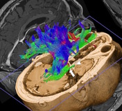
Image 3 MRI ( create )
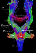
Image 4 MRI (Tractography) ( update )

Image 5 MRI ( create )

Image 6 MRI ( create )


 Unable to process the form. Check for errors and try again.