
Extensor digitorum brevis manus muscle
Updates to Article Attributes
The extensor digitorum brevis manus (EDBM) muscle is an accessory muscle in the hand and is a normal anatomical variant.
Epidemiology
It is thought to be present in ~ 3% of the population 1. It is often painless although rarely can present as a painful mass over the dorsal aspect of the hand. It can be bilateral in up to half of cases.
Gross anatomy
The EDBM muscle lies along the ulnar side of the extensor tendon of the index finger (usually fourth wrist compartment 5). It commonly arises at the distal end of the radius and posterior radiocarpal ligament to insert, most commonly on the index finger. However, insertion can also be seen on the 3rd, 4th, or 5th digits as well as multiple insertions on more than one digit.
Blood supply
- arterial supply - posterior interosseous nerve
Nerve supply
Clinical presentation
The muscle is usually painless but may occasionally be associated with exercise-induced pain or tenosynovitis of the extensor tendons.
Radiographic features
Plain film
Can be normal
Ultrasound
Sonography may reveal a soft-tissue mass with a musclelike echo texture, on real time this usually undergoes morphologic changes during active finger extension.
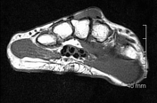
MRI
Signal characteristics include
- T1/T2/PD - Iso dense to muscle on all sequences
- C+ (Gd) - no enhancement in uncomplicated cases (e.g. unless there is any inflammation etc.)
Summary
- origin - distal radius and posterior radiocarpal ligmanet
- insertion - extensor hood of index or middle fingers (variable)
- innervation - posterior interosseous nerve
- action - extension of the fingers
-<p>The <strong>extensor digitorum brevis manus (EDBM)</strong> muscle is an <a href="/articles/accessory-muscle" title="accessory muscle">accessory muscle</a> in the hand and is a normal anatomical variant. </p><h4>Epidemiology</h4><p>It is thought to be present in ~ 3% of the population <sup>1</sup>. It is often painless although rarely can present as a painful mass over the dorsal aspect of the hand. It can be bilateral in up to half of cases.</p><h4>Gross anatomy</h4><p>The EDBM muscle lies along the ulnar side of the extensor tendon of the index finger (usually fourth wrist compartment <sup>5</sup>). It commonly arises at the distal end of the radius and posterior radiocarpal ligament to insert, most commonly on the index finger. However, insertion can also be seen on the 3<sup>rd</sup>, 4<sup>th</sup>, or 5<sup>th</sup> digits as well as multiple insertions on more than one digit. </p><h4>Blood supply</h4><ul><li>arterial supply - <a href="/articles/posterior-interosseous-nerve" title="posterior interosseous nerve">posterior interosseous nerve</a>-</li></ul><h4>Nerve supply</h4><ul><li><a href="/articles/posterior-interosseous-nerve" title="posterior interosseous nerve">posterior interosseous nerve</a></li></ul><h4>Clinical presentation</h4><p>The muscle is usually painless but may occasionally be associated with exercise-induced pain or tenosynovitis of the extensor tendons.</p><h4>Radiographic features</h4><h5>Plain film</h5><p>Can be normal</p><h5>Ultrasound</h5><p>Sonography may reveal a soft-tissue mass with a musclelike echo texture, on real time this usually undergoes morphologic changes during active finger extension.</p><h5>MRI</h5><p>Signal characteristics include</p><ul>-<li>-<strong>T1/T2/PD</strong> - Iso dense to muscle on all sequences</li>-<li>-<strong>C+ (Gd) </strong>- no enhancement in uncomplicated cases (e.g. unless there is any inflammation etc.)</li>- +<p>The <strong>extensor digitorum brevis manus (EDBM)</strong> muscle is an <a href="/articles/accessory-muscle">accessory muscle</a> in the hand and is a normal anatomical variant. </p><h4>Epidemiology</h4><p>It is thought to be present in ~ 3% of the population <sup>1</sup>. It is often painless although rarely can present as a painful mass over the dorsal aspect of the hand. It can be bilateral in up to half of cases.</p><h4>Gross anatomy</h4><p>The EDBM muscle lies along the ulnar side of the extensor tendon of the index finger (usually fourth wrist compartment <sup>5</sup>). It commonly arises at the distal end of the radius and posterior radiocarpal ligament to insert, most commonly on the index finger. However, insertion can also be seen on the 3<sup>rd</sup>, 4<sup>th</sup>, or 5<sup>th</sup> digits as well as multiple insertions on more than one digit. </p><h4>Blood supply</h4><ul><li>arterial supply - <a href="/articles/posterior-interosseous-nerve">posterior interosseous nerve</a>
- +</li></ul><h4>Nerve supply</h4><ul><li><a href="/articles/posterior-interosseous-nerve">posterior interosseous nerve</a></li></ul><h4>Clinical presentation</h4><p>The muscle is usually painless but may occasionally be associated with exercise-induced pain or tenosynovitis of the extensor tendons.</p><h4>Radiographic features</h4><h5>Plain film</h5><p>Can be normal</p><h5>Ultrasound</h5><p>Sonography may reveal a soft-tissue mass with a musclelike echo texture, on real time this usually undergoes morphologic changes during active finger extension.</p><h5>MRI</h5><p>Signal characteristics include</p><ul>
- +<li>
- +<strong>T1/T2/PD</strong> - Iso dense to muscle on all sequences</li>
- +<li>
- +<strong>C+ (Gd) </strong>- no enhancement in uncomplicated cases (e.g. unless there is any inflammation etc.)</li>
-<li>-<strong>origin</strong> - distal radius and posterior radiocarpal ligmanet</li>-<li>-<strong>insertion</strong> - extensor hood of index or middle fingers (variable)</li>-<li>-<strong>innervation</strong> - posterior interosseous nerve</li>-<li>-<strong>action</strong> - extension of the fingers</li>- +<li>
- +<strong>origin</strong> - distal radius and posterior radiocarpal ligmanet</li>
- +<li>
- +<strong>insertion</strong> - extensor hood of index or middle fingers (variable)</li>
- +<li>
- +<strong>innervation</strong> - posterior interosseous nerve</li>
- +<li>
- +<strong>action</strong> - extension of the fingers</li>
References changed:
- 4. Sookur PA, Naraghi AM, Bleakney RR et-al. Accessory muscles: anatomy, symptoms, and radiologic evaluation. Radiographics. 2008;28 (2): 481-99. <a href="http://radiographics.rsna.org/content/28/2/481.full">Radiographics (full text)</a> - <a href="http://dx.doi.org/10.1148/rg.282075064">doi:10.1148/rg.282075064</a> - <a href="http://www.ncbi.nlm.nih.gov/pubmed/18349452">Pubmed citation</a><span class="ref_v3"></span>
- 6. Rodríguez-Niedenführ M, Vázquez T, Golanó P et-al. Extensor digitorum brevis manus: anatomical, radiological and clinical relevance. A review. Clin Anat. 2002;15 (4): 286-92. <a href="http://onlinelibrary.wiley.com/doi/10.1002/ca.10027/abstract">Clin Anat (abstract)</a> - <a href="http://dx.doi.org/10.1002/ca.10027">doi:10.1002/ca.10027</a> - <a href="http://www.ncbi.nlm.nih.gov/pubmed/12112357">Pubmed citation</a><span class="ref_v3"></span>
- 1. Ouellette H, Thomas B, Torriani M. Using Dynamic Sonography to Diagnose Extensor Digitorum Brevis Manus. AJR Am J Roentgenol. 2003;181(5):1224-6. <a href="https://doi.org/10.2214/ajr.181.5.1811224">doi:10.2214/ajr.181.5.1811224</a> - <a href="https://www.ncbi.nlm.nih.gov/pubmed/14573408">Pubmed</a>
- 2. Anderson M, Benedetti P, Walter J, Steinberg D. MR Appearance of the Extensor Digitorum Manus Brevis Muscle: A Pseudotumor of the Hand. AJR Am J Roentgenol. 1995;164(6):1477-9. <a href="https://doi.org/10.2214/ajr.164.6.7754896">doi:10.2214/ajr.164.6.7754896</a> - <a href="https://www.ncbi.nlm.nih.gov/pubmed/7754896">Pubmed</a>
- 3. Bianchi S, Della Santa D, Glauser T et-al. Sonography of masses of the wrist and hand. AJR Am J Roentgenol. 2008;191 (6): 1767-75. <a href="http://dx.doi.org/10.2214/AJR.07.4003">doi:10.2214/AJR.07.4003</a> - <a href="http://www.ncbi.nlm.nih.gov/pubmed/19020249">Pubmed citation</a><span class="ref_v3"></span>
- 5. Froelich JM, Bidgoli-Moghaddam M, Moran SL. Bilateral extensor digitorum brevis manus. Orthopedics. 2012;35 (9): e1431-3. <a href="http://dx.doi.org/10.3928/01477447-20120822-34">doi:10.3928/01477447-20120822-34</a> - <a href="http://www.ncbi.nlm.nih.gov/pubmed/22955414">Pubmed citation</a><span class="ref_v3"></span>
- 4- Sookur PA, Naraghi AM, Bleakney RR et-al. Accessory muscles: anatomy, symptoms, and radiologic evaluation. Radiographics. 2008;28 (2): 481-99. <a href="http://radiographics.rsna.org/content/28/2/481.full">Radiographics (full text)</a> - <a href="http://dx.doi.org/10.1148/rg.282075064">doi:10.1148/rg.282075064</a> - <a href="http://www.ncbi.nlm.nih.gov/pubmed/18349452">Pubmed citation</a><span class="ref_v3"></span>
- 6- Rodríguez-Niedenführ M, Vázquez T, Golanó P et-al. Extensor digitorum brevis manus: anatomical, radiological and clinical relevance. A review. Clin Anat. 2002;15 (4): 286-92. <a href="http://onlinelibrary.wiley.com/doi/10.1002/ca.10027/abstract">Clin Anat (abstract)</a> - <a href="http://dx.doi.org/10.1002/ca.10027">doi:10.1002/ca.10027</a> - <a href="http://www.ncbi.nlm.nih.gov/pubmed/12112357">Pubmed citation</a><span class="ref_v3"></span>
- 1- Ouellette H, Thomas BJ, Torriani M. Using dynamic sonography to diagnose extensor digitorum brevis manus. AJR Am J Roentgenol. 2003;181 (5): 1224-6. <a href="http://dx.doi.org/10.2214/ajr.181.5.1811224">doi:10.2214/ajr.181.5.1811224</a> - <a href="http://www.ncbi.nlm.nih.gov/pubmed/14573408">Pubmed citation</a><span class="ref_v3"></span>
- 2- Anderson MW, Benedetti P, Walter J et-al. MR appearance of the extensor digitorum manus brevis muscle: a pseudotumor of the hand. AJR Am J Roentgenol. 1995;164 (6): 1477-9. <a href="http://dx.doi.org/10.2214/ajr.164.6.7754896">doi:10.2214/ajr.164.6.7754896</a> - <a href="http://www.ncbi.nlm.nih.gov/pubmed/7754896">Pubmed citation</a><span class="ref_v3"></span>
- 3- Bianchi S, Della Santa D, Glauser T et-al. Sonography of masses of the wrist and hand. AJR Am J Roentgenol. 2008;191 (6): 1767-75. <a href="http://dx.doi.org/10.2214/AJR.07.4003">doi:10.2214/AJR.07.4003</a> - <a href="http://www.ncbi.nlm.nih.gov/pubmed/19020249">Pubmed citation</a><span class="ref_v3"></span>
- 5- Froelich JM, Bidgoli-Moghaddam M, Moran SL. Bilateral extensor digitorum brevis manus. Orthopedics. 2012;35 (9): e1431-3. <a href="http://dx.doi.org/10.3928/01477447-20120822-34">doi:10.3928/01477447-20120822-34</a> - <a href="http://www.ncbi.nlm.nih.gov/pubmed/22955414">Pubmed citation</a><span class="ref_v3"></span>
Image 1 MRI (T1) ( create )


 Unable to process the form. Check for errors and try again.