
Foot radiograph (an approach)
Updates to Article Attributes
Body
was changed:
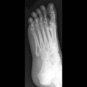
Foot radiographs are commonly performed in Emergency departments, usually after sport-related trauma and often with a clinical request that states lateral border pain. Remember to check the whole film though. Often, a foot x-ray is also requested for the investigation of osteomyelitis, arthritides, or a bone lesion.
This article relates mainly to the traumatic injuries to the foot.
A basic review should start with AP and lateral views (including the entire foot and ankle). With the exception of the trauma, these views should be acquired with weight bearing if the patient can tolerate.
Systematic review
Lisfranc complex
The Lisfranc joint is hugely important for stability. Injury to it may be subtle and if missed, disastrous.
- medial borders of
2nd2nd metatarsal and intermediate cuneiform should line up on the DP (dorsiplantar) view - medial borders of
3rd3rd metatarsal and lateral cuneiform should line up on the oblique view - if there is any step in either line, think Lisfranc injury
Medial aspect (DP)
-
1st1st and2nd2nd metatarsals - medial and intermediate cuneiform
Lateral aspect (oblique)
-
3rd3rd,4th4th and5th5th metatarsals - lateral cuneiform
- navicular and cuboid
Bone review
- check around the cortex of every bone
- start proximally and work distally, medial to lateral
- check any tarsal coalition
- look for any bone that isn't attached
- is it an ossicle, an avulsion or bone fragment?
- don't call normal variant anatomy a fracture!
- don't call an unfused base of 5th apophysis a fracture!
Common pathology
Lisfranc injury
- Lisfranc ligament between
1st1st and2nd2nd metatarsal bases - the ligament stabilises the foot
- widening of the
1st/2nd1st/2nd metatarsal space - a line along the medial margins of the
2nd2nd metatarsal and intermediate cuneiform will be irregular - disruption suggests a huge injury
- usually a crush injury or axial load to a plantarflexed foot
- more: Lisfranc injury
Chopart injury
- fracture / dislocation of the mid-tarsal joint (Chopart joint) of the foot, i.e. talonavicular and calcaneocuboid joints
- foot is usually dislocated medially and superiorly as it is plantar flexed and inverted, usually as a result of high energy impact
- more: Chopart fracture
Avulsion fractures associated with ankle sprain:
Avulsion of the 5th5th metatarsal styloid
- 90% of base of
5th5th metatarsal fractures - avulsion of peroneus brevis tendon
- forced inversion of plantarflexed foot (tennis fracture)
- transverse fracture through tuberosity extending to tarsometatarsal joint
- excellent prognosis
- more: 5th metatarsal styloid avulsion
Dorsal capsular avulsion
- curvilinear calcification dorsal to talar head or navicular bone
Extensor digitorum brevis avulsion fracture
- thin calcification adjacent to anterolateral calcaneus on oblique view
Snowboarder's fracture
Jones fracture
- base of 5th metatarsal fracture
- transverse fracture 1.5-2 cm from tip of proximal tuberosity
- forced inversion of plantarflexed foot
- transverse fracture through diaphysis
- high risk of nonunion
- more: Jones fracture
Calcaneal fracture
- calcaneal tuberosity avulsion fracture
- extra-articular lover fracture (or Casanova fracture)
- intra-articular lover fracture
- calcaneal stress fracture
Don't miss
Stress fracture
- commonly affect
2nd2nd and3rd3rd metatarsal shafts - abnormal stresses lead to microfractures, e.g. marching
- look for transverse fracture, periosteal reaction or callus
- more: metatarsal stress fracture
-<li>medial borders of 2nd metatarsal and intermediate cuneiform should line up on the DP (dorsiplantar) view</li>-<li>medial borders of 3rd metatarsal and lateral cuneiform should line up on the oblique view</li>- +<li>medial borders of 2<sup>nd</sup> metatarsal and intermediate cuneiform should line up on the DP (dorsiplantar) view</li>
- +<li>medial borders of 3<sup>rd</sup> metatarsal and lateral cuneiform should line up on the oblique view</li>
-<li>1st and 2nd metatarsals</li>- +<li>1<sup>st</sup> and 2<sup>nd</sup> metatarsals</li>
-<li>3rd, 4th and 5th metatarsals</li>- +<li>3<sup>rd</sup>, 4<sup>th</sup> and 5<sup>th </sup>metatarsals</li>
-<li>Lisfranc ligament between 1st and 2nd metatarsal bases</li>- +<li>Lisfranc ligament between 1<sup>st</sup> and 2<sup>nd</sup> metatarsal bases</li>
-<li>widening of the 1st/2nd metatarsal space</li>-<li>a line along the medial margins of the 2nd metatarsal and intermediate cuneiform will be irregular</li>- +<li>widening of the 1<sup>st</sup>/2<sup>nd</sup> metatarsal space</li>
- +<li>a line along the medial margins of the 2<sup>nd</sup> metatarsal and intermediate cuneiform will be irregular</li>
-</ul><h5>Avulsion fractures associated with ankle sprain:</h5><h6>Avulsion of the 5th metatarsal styloid</h6><ul>-<li>90% of base of 5th metatarsal fractures</li>- +</ul><h5>Avulsion fractures associated with ankle sprain:</h5><h6>Avulsion of the 5<sup>th</sup> metatarsal styloid</h6><ul>
- +<li>90% of base of 5<sup>th</sup> metatarsal fractures</li>
-<li>commonly affect 2nd and 3rd metatarsal shafts</li>- +<li>commonly affect 2<sup>nd</sup> and 3<sup>rd </sup>metatarsal shafts</li>
Images Changes:
Image 4 X-ray (Frontal) ( update )

Caption
was changed:
Image 5 X-ray (Oblique) ( update )
Caption
was changed:
Image 7 Annotated image (Phalanges) ( update )

Caption
was changed:
Figure 3: DP phalanges
Image 8 Annotated image (Metatarsals) ( update )

Caption
was changed:
Figure 4: DP metatarsals
Image 9 Annotated image (Lisfranc) ( update )

Caption
was changed:
Figure 5: DP Lisfranc
Image 10 Annotated image (Tarsals) ( update )

Caption
was changed:
Figure 6: DP tarsals
Image 11 Annotated image (Oblique) ( update )

Caption
was changed:
Image 12 Annotated image (Oblique) ( update )

Caption
was changed:
Image 13 Annotated image (Oblique) ( update )

Caption
was changed:
Image 14 Annotated image (Oblique) ( update )

Caption
was changed:
Image 15 Annotated image (Lateral projection.) ( update )

Caption
was changed:


 Unable to process the form. Check for errors and try again.