
Honeycombing (lungs)
Updates to Article Attributes
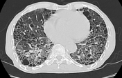
Honeycombing is a CT imaging descriptor referring to clustered cystic air spaces (between 3-10 mm in diameter, but occasionally as large as 2.5 cm) that are usually subpleural, peripheral and basal in distribution. They can be subdivided into:
The walls of the cysts are well-defined and often thick (1-3 mm) 4. They represent an irreversible finding commonly seen in diffuse pulmonary fibrosis (usual interstitial pneumonia UIP).
Pathology
Honeycombing consists of enlarged air spaces with thick fibrotic walls lined by bronchiolar epithelium and often filled with mucin and inflammatory cells 6.
History and etymology
The term “honeycomb lung” is thought to have originated in the 19th century in Germany and is thought to have first appeared in 1949 in a study by N Oswald and T Parkinson 5.
Differential diagnosis
- in some situations
- airspace consolidation in the presence of pulmonary emphysema can mimic this appearance
-<li><a title="Bronchiolectasis" href="/articles/bronchiolectasis-1">traction bronchiolectasis</a></li>- +<li><a href="/articles/bronchiolectasis-1">traction bronchiolectasis</a></li>
-<a href="/articles/air-space-opacification-1">airspace consolidation</a> in the presence of <a title="Pulmonary emphysema" href="/articles/pulmonary-emphysema">pulmonary emphysema</a> can mimic this appearance</li>- +<a href="/articles/air-space-opacification-1">airspace consolidation</a> in the presence of <a href="/articles/pulmonary-emphysema">pulmonary emphysema</a> can mimic this appearance</li>
Image 6 CT (lung window) ( create )


 Unable to process the form. Check for errors and try again.