
Hydrocele
Updates to Article Attributes
A hydroceleHydrocele is an acquired or congenital serous fluid collection within layers of the tunicas vaginalistunica vaginalis 1. It is the most common form of testicular enlargement 2.
Clinical presentation
Most hydrocele are acquired and present with progressing painless scrotal mass. Characteristically, hydrocele transilluminates when evaluted with light source during physical examination. However, hydroceles can be secondarily infected (see pyocele).
Epidemiology
Hydrocele can be diagnosed at any age, with congenital hydrocele being more common in children.
Pathology
Congenital
There are two subtypes of congenital hydrocele 1-2 :
- encysted type with no communication with the peritoneum or tunica vaginalis, also called spermatic cord cyst.
- funicular type which communicates with the peritoneum at the internal ring and doesn't surround the testis. This type is also called funiculocele. They are more frequently encoutered in children and premature infant 2.
Acquired aetiology
- trauma
- epididymitis
- testicular torsion
- testicular neoplasm
Radiographic features
Ultrasound
Ultrasound is the first modality usually used to evaluate hydroceles. It presents as a simple fluid collection surrounding the testits. It is avascular on Doppler evaluation. It may contain septations, calcifications and cholesterol 2.
A funiculocele is a sub type of hydrocele, however, it doesn't surround the testis. They can also appear larger with straining (valsalva) 2. It may contain fibrous adhesions, giving a beaded appearance to the spermatic cord (pachyvaginalitis) 3.
The encysted subtype shows no communication with the peritoneum and it usually only involves the spermatic cord.
MRI
On MRI, signal characteristics of the hydroceles are :
-
T1:
- lowlow signal -
T2:
- highhigh signal
This represents the simple serous fluid component of the hydrocele.
Prognosis
In infants most hydroceles (around 90%) resolve spontaneously and their are thought to result in incomplete obliteration of the processus vaginalis 4. It is important to assess for any associated herniations in these patients.
Differential diagnosis
Imaging differential considerations include
Rarely, a scrotal tunica cyst and a scrotal mesothelioma can look like hydrocele. It is usually simple to distinguish them from hydroceles.
See also
-<p>A <strong>hydrocele</strong> is an acquired or congenital serous fluid collection within layers of the tunicas vaginalis <sup>1</sup>. It is the most common form of testicular enlargement <sup>2</sup>.</p><h4>Clinical presentation</h4><p>Most hydrocele are acquired and present with progressing painless <a title="scrotal mass" href="/articles/scrotal-mass">scrotal mass</a>. Characteristically, hydrocele transilluminates when evaluted with light source during physical examination. However, hydroceles can be secondarily infected (see <a title="pyocele" href="/articles/pyocele">pyocele</a>).</p><h4>Epidemiology</h4><p>Hydrocele can be diagnosed at any age, with congenital hydrocele being more common in children.</p><h4>Pathology</h4><h6>Congenital</h6><p>There are two subtypes of congenital hydrocele <sup>1-2</sup> :</p><ul>-<li>encysted type with no communication with the peritoneum or tunica vaginalis, also called <a href="/articles/spermatic-cord-cyst" title="spermatic cord cyst">spermatic cord cyst</a>.</li>-<li>funicular type which communicates with the peritoneum at the internal ring and doesn't surround the testis. This type is also called funiculocele. They are more frequently encoutered in children and premature infant <sup>2</sup>.</li>- +<p><strong>Hydrocele</strong> is an acquired or congenital serous fluid collection within layers of the tunica vaginalis <sup>1</sup>. It is the most common form of testicular enlargement <sup>2</sup>.</p><h4>Clinical presentation</h4><p>Most hydrocele are acquired and present with progressing painless <a href="/articles/scrotal-mass">scrotal mass</a>. Characteristically, hydrocele transilluminates when evaluted with light source during physical examination. However, hydroceles can be secondarily infected (see <a href="/articles/pyocele">pyocele</a>).</p><h4>Epidemiology</h4><p>Hydrocele can be diagnosed at any age, with congenital hydrocele being more common in children.</p><h4>Pathology</h4><h6>Congenital</h6><p>There are two subtypes of congenital hydrocele <sup>1-2</sup> :</p><ul>
- +<li>encysted type with no communication with the peritoneum or tunica vaginalis, also called <a href="/articles/spermatic-cord-cyst">spermatic cord cyst</a>.</li>
- +<li>funicular type which communicates with the peritoneum at the internal ring and doesn't surround the testis. This type is also called funiculocele. They are more frequently encoutered in children and premature infant <sup>2</sup>.</li>
-<li>trauma</li>-<li><a title="Epididymitis" href="/articles/epididymitis">epididymitis</a></li>-<li><a title="Testicular torsion" href="/articles/testicular-torsion">testicular torsion</a></li>-<li>testicular neoplasm</li>-</ul><h4>Radiographic features</h4><h5>Ultrasound</h5><p>Ultrasound is the first modality usually used to evaluate hydroceles. It presents as a simple fluid collection surrounding the testits. It is avascular on Doppler evaluation. It may contain septations, calcifications and cholesterol <sup>2</sup>.</p><p>A <a title="funiculocele" href="/articles/funiculocele">funiculocele</a> is a sub type of hydrocele, however, it doesn't surround the testis. They can also appear larger with straining (valsalva) <sup>2</sup>. It may contain fibrous adhesions, giving a beaded appearance to the spermatic cord (pachyvaginalitis) <sup>3.</sup></p><p>The encysted subtype shows no communication with the peritoneum and it usually only involves the spermatic cord.</p><h5>MRI</h5><p>On MRI, signal characteristics of the hydroceles are :</p><ul>-<li>-<strong>T1</strong> - low signal</li>-<li>-<strong>T2</strong> - high signal</li>-</ul><p>This represents the simple serous fluid component of the hydrocele.</p><h4>Differential diagnosis</h4><p>Imaging differential considerations include</p><ul>-<li><a title="Spermatocele" href="/articles/spermatocoele">spermatocele</a></li>-<li><a title="Pyocele" href="/articles/pyocele">pyocele</a></li>-<li><a title="Hematocele" href="/articles/hematocele">haematocele</a></li>-<li><a title="Abdominal hernias" href="/articles/abdominal-hernia">inguinoscrotal hernia</a></li>-</ul><p>Rarely, a <a title="scrotal tunica cyst " href="/articles/scrotal-tunica-cyst">scrotal tunica cyst </a>and a <a title="scrotal mesothelioma " href="/articles/scrotal-mesothelioma">scrotal mesothelioma </a>can look like hydrocele. It is usually simple to distinguish them from hydroceles.</p><h4>See also</h4><ul>-<li><a href="/articles/hydrocoele-of-the-canal-of-nuck" title="Hydrocoele of canal of Nuck">hydrocoele of canal of Nuck</a></li>-<li><a href="/articles/spermatic-cord-hydrocele" title="Spermatic cord hydrocoele">spermatic cord hydrocoele</a></li>- +<li>trauma</li>
- +<li><a href="/articles/epididymitis">epididymitis</a></li>
- +<li><a href="/articles/testicular-torsion">testicular torsion</a></li>
- +<li>testicular neoplasm</li>
- +</ul><h4>Radiographic features</h4><h5>Ultrasound</h5><p>Ultrasound is the first modality usually used to evaluate hydroceles. It presents as a simple fluid collection surrounding the testits. It is avascular on Doppler evaluation. It may contain septations, calcifications and cholesterol <sup>2</sup>.</p><p>A <a href="/articles/funiculocele">funiculocele</a> is a sub type of hydrocele, however, it doesn't surround the testis. They can also appear larger with straining (valsalva) <sup>2</sup>. It may contain fibrous adhesions, giving a beaded appearance to the spermatic cord (pachyvaginalitis) <sup>3.</sup></p><p>The encysted subtype shows no communication with the peritoneum and it usually only involves the spermatic cord.</p><h5>MRI</h5><p>On MRI, signal characteristics of the hydroceles are :</p><ul>
- +<li>
- +<strong>T1:</strong> low signal</li>
- +<li>
- +<strong>T2:</strong> high signal</li>
- +</ul><p>This represents the simple serous fluid component of the hydrocele.</p><h4>Prognosis</h4><p>In infants most hydroceles (around 90%) resolve spontaneously and their are thought to result in incomplete obliteration of the processus vaginalis<sup> 4</sup>. It is important to assess for any associated herniations in these patients.</p><h4>Differential diagnosis</h4><p>Imaging differential considerations include</p><ul>
- +<li><a href="/articles/spermatocoele">spermatocele</a></li>
- +<li><a href="/articles/pyocele">pyocele</a></li>
- +<li><a href="/articles/hematocele">haematocele</a></li>
- +<li><a href="/articles/abdominal-hernia">inguinoscrotal hernia</a></li>
- +</ul><p>Rarely, a <a href="/articles/scrotal-tunica-cyst">scrotal tunica cyst </a>and a <a href="/articles/scrotal-mesothelioma">scrotal mesothelioma </a>can look like hydrocele. It is usually simple to distinguish them from hydroceles.</p><h4>See also</h4><ul>
- +<li><a href="/articles/hydrocoele-of-the-canal-of-nuck">hydrocoele of canal of Nuck</a></li>
- +<li><a href="/articles/spermatic-cord-hydrocele">spermatic cord hydrocoele</a></li>
References changed:
- 4. Naji H, Ingolfsson I, Isacson D, Svensson J. Decision Making in the Management of Hydroceles in Infants and Children. Eur J Pediatr. 2012;171(5):807-10. <a href="https://doi.org/10.1007/s00431-011-1628-x">doi:10.1007/s00431-011-1628-x</a> - <a href="https://www.ncbi.nlm.nih.gov/pubmed/22105873">Pubmed</a>
- 5. Christensen T, Cartwright P, Devries C, Snow B. New Onset of Hydroceles in Boys over 1 Year of Age. Int J Urol. 2006;13(11):1425-7. <a href="https://doi.org/10.1111/j.1442-2042.2006.01583.x">doi:10.1111/j.1442-2042.2006.01583.x</a> - <a href="https://www.ncbi.nlm.nih.gov/pubmed/17083397">Pubmed</a>
Image ( update )
Image 1 MRI (T2) ( update )

Image 2 Ultrasound ( update )

Image 3 Ultrasound ( update )

Image 5 Ultrasound (Transverse) ( update )
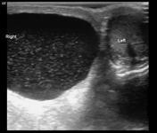
Image 8 ( update )


 Unable to process the form. Check for errors and try again.