
Hyoid bone
Updates to Article Attributes
The hyoid is a "horseshoe"-shaped bone that serves as a structural anchor in the mid-neck. It is a place of convergence of multiple small neck muscles that permit the pharyngeal phase of swallowing. Structures in the neck are often located in relation to the hyoid (i.e. suprahyoid neck; infrahyoid neck).
Summary
-
location: midline neck, anterior to the trachea
- the level of the hyoid separates levels II from III in the lateral neck and levels Ia and VI in the anterior neck
- articulations: none
- blood supply and innervation: branches of the external carotid artery
- relations: numerous muscles insert on the hyoid, detailed below
Gross anatomy
Location
MidThe hyoid bone is located in the mid-neck, above the thyroid cartilage, anterior to the trachea. Structures in the neck are often located in relation to the hyoid (e.g. suprahyoid or infrahyoid neck)
The position of the hyoid defines a few lymph node levels of the neck. Anteriorly, the hyoid separates levels Ia (above) from VI (below). Laterally, the level of the hyoid separates level II from level III.
Blood supply
Branches of the external carotid artery, predominately the infrahyoid artery off the superior thyroid artery and branches off the lingual artery.
Relations
The hyoid consists of three parts:
- a body
- two superior genu
- two inferior genu
Attachments
Multiple small neck muscles insert into these regionsin the three parts:
- body
- superior genu
- inferior genu
-
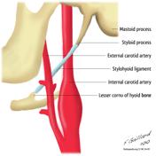
no muscles, just thestylohyoid ligament (i.e. no muscular attachment)
-
Relations
The position of the hyoid defines a few lymph node levels of the neck. Anteriorly, the hyoid separates levels Ia (above) from VI (below). Laterally, the level of the hyoid separates level II from level III.
Blood supply
Branches of the external carotid artery, predominantly the infrahyoid artery off the superior thyroid artery and branches off the lingual artery.
Radiographic features
The hyoid is the ossified horseshoe-shaped structure just superior to the thyroid cartilage.
Related pathology
Movement of the hyoid (hyoid elevation) is an indication of the strength of the pharyngeal muscles during a modified barium swallow study.
-<p>The <strong>hyoid </strong>is a "horseshoe"-shaped bone that serves as a structural anchor in the mid-neck. It is a place of convergence of multiple small neck muscles that permit the pharyngeal phase of swallowing.</p><h4>Summary</h4><ul>- +<p>The <strong>hyoid </strong>is a "horseshoe"-shaped bone that serves as a structural anchor in the mid-neck. It is a place of convergence of multiple small neck muscles that permit the pharyngeal phase of swallowing. Structures in the neck are often located in relation to the hyoid (i.e. suprahyoid neck; infrahyoid neck). </p><h4>Summary</h4><ul>
-</ul><h4>Gross anatomy</h4><h5>Location</h5><p>Mid-neck, above the thyroid cartilage, anterior to the trachea. Structures in the neck are often located in relation to the hyoid (e.g. suprahyoid or infrahyoid neck)</p><p>The position of the hyoid defines a few <a href="/articles/lymph-node-levels-of-the-neck">lymph node levels of the neck</a>. Anteriorly, the hyoid separates levels Ia (above) from VI (below). Laterally, the level of the hyoid separates level II from level III.</p><h5>Blood supply </h5><p>Branches of the <a href="/articles/external-carotid-artery-1">external carotid artery</a>, predominately the <a href="/articles/infrahyoid-artery">infrahyoid artery</a> off the <a href="/articles/superior-thyroid-artery">superior thyroid artery</a> and branches off the <a href="/articles/lingual-artery">lingual artery</a>.</p><h5>Relations</h5><p>The hyoid consists of three parts:</p><ul>- +</ul><h4>Gross anatomy</h4><p>The hyoid bone is located in the mid-neck, above the <a title="thyroid cartilage" href="/articles/thyroid-cartilage">thyroid cartilage</a>, anterior to the <a title="trachea" href="/articles/trachea">trachea</a>. The hyoid consists of three parts:</p><ul>
-</ul><p>Multiple small neck muscles insert into these regions:</p><ul>- +</ul><h5>Attachments</h5><p>Multiple small neck muscles insert in the three parts:</p><ul>
-<li>inferior genu<ul><li>no muscles, just the <a href="/articles/stylohyoid-ligament">stylohyoid ligament</a>-</li></ul>- +<li>inferior genu<ul><li>
- +<a href="/articles/stylohyoid-ligament">stylohyoid ligament</a> (i.e. no muscular attachment)</li></ul>
-</ul><h4>Radiographic features</h4><p>The hyoid is the ossified horseshoe-shaped structure just superior to the thyroid cartilage.</p><h4>Related pathology</h4><p>Movement of the hyoid (<a href="/articles/hyoid-elevation">hyoid elevation</a>) is an indication of the strength of the pharyngeal muscles during a <a href="/articles/modified-barium-swallow">modified barium swallow</a> study.</p>- +</ul><h5>Relations</h5><p>The position of the hyoid defines a few <a href="/articles/lymph-node-levels-of-the-neck">lymph node levels of the neck</a>. Anteriorly, the hyoid separates levels Ia (above) from VI (below). Laterally, the level of the hyoid separates level II from level III.</p><h4>Blood supply </h4><p>Branches of the <a href="/articles/external-carotid-artery-1">external carotid artery</a>, predominantly the <a href="/articles/infrahyoid-artery">infrahyoid artery</a> off the <a href="/articles/superior-thyroid-artery">superior thyroid artery</a> and branches off the <a href="/articles/lingual-artery">lingual artery</a>.</p><h4>Radiographic features</h4><p>The hyoid is the ossified horseshoe-shaped structure just superior to the thyroid cartilage.</p><h4>Related pathology</h4><p>Movement of the hyoid (<a href="/articles/hyoid-elevation">hyoid elevation</a>) is an indication of the strength of the pharyngeal muscles during a <a href="/articles/modified-barium-swallow">modified barium swallow</a> study.</p>
Image 3 Diagram ( update )


 Unable to process the form. Check for errors and try again.