
Hysterosalpingogram
Updates to Article Attributes
Body
was changed:
Hysterosalpingogram (HSG) is a fluoroscopic examination of the uterus and the Fallopian tubes, most commonly used in the investigation of infertility or recurrent spontaneous abortions.
Technique
- the procedure should be performed during the proliferative phase of the patient’s menstrual cycle (days 6-12), when the endometrium is thinnest
- this improves visualisation of the uterine cavity, and also minimises the possibility that the patient may be pregnant 1
- if there is any uncertainty about the patient’s pregnancy status, a bHCG is warranted prior to commencing.
- after an antiseptic clean of the external genital area, a vaginal speculum is inserted with the patient in the lithotomy position ; the cervix is cleaned with an aseptic solution.
- catheterisation of the cervix is then performed ; the type of device used depends on local practice preferences
- e.g. 6 Fr Foley catheter with balloon inflation, or
- any one of a range of available HSG catheters or metal cannulas 3.
- whatever the device, it should be primed with contrast prior to commencing to avoid the introduction of gas bubbles which may provide a false positive appearance of a filling defect.
- water soluble iodinated contrast is subsequently injected slowly under fluoroscopic guidance.
- Some radiologists use iodinated oil (Lipiodol) as contrast when the indication is for lack of fertility. Some authors report increased fertility after its use. This remain controversial however 8.
- a typical fluoroscopic examination includes preliminary frontal view of the pelvis, as well as subsequent spot images that demonstrate uterine endometrial contour, filled fallopian tubes and bilateral intraperitoneal spill of contrast, to establish tubal patency.
Contraindications
- pregnancy
- active pelvic infection
- recent uterine or tubal surgery
Complications
Common but self limiting
- abdominal cramping
- PV spotting
Rare but serious
- pelvic infection
- contrast reaction
Detectable pathology
Conditions which may be detected with HSG include:
Uterine
- uterine congenital anomalies
- submucosal uterine fibroids
- uterine malignancy
- adenomyosis
- intrauterine adhesions
- uterine (endometrial) polyps
Tubal
-
obliteration of fallopian tubes : usually secondary to previous pelvic inflammation. It must be differentiated from incomplete tubal opacification due to tubal spasm, or underfilling of the uterus with contrast 2
. -
tubal polyps 6
. - tubal malignancy
- hydrosalpinx
-
salpingitis isthmica nodosa (SIN) 4
. - tubal spasm 6: can be physiological
-<a href="/articles/obliteration-of-fallopian-tubes">obliteration of fallopian tubes</a> : usually secondary to previous pelvic inflammation. It must be differentiated from incomplete tubal opacification due to tubal spasm, or underfilling of the uterus with contrast <sup>2</sup>.</li>- +<a href="/articles/obliteration-of-fallopian-tubes">obliteration of fallopian tubes</a> : usually secondary to previous pelvic inflammation. It must be differentiated from incomplete tubal opacification due to tubal spasm, or underfilling of the uterus with contrast <sup>2</sup>
- +</li>
-<a href="/articles/tubal-polyps">tubal polyps</a> <sup>6</sup>.</li>- +<a href="/articles/tubal-polyps">tubal polyps</a> <sup>6</sup>
- +</li>
-<a href="/articles/salpingitis-isthmica-nodosa-1">salpingitis isthmica nodosa</a> (SIN) <sup>4</sup>.</li>- +<a href="/articles/salpingitis-isthmica-nodosa-1">salpingitis isthmica nodosa</a> (SIN) <sup>4</sup>
- +</li>
-<a href="/articles/tubal-spasm">tubal spasm</a> <sup>6</sup> : can be physiological</li>- +<a href="/articles/tubal-spasm">tubal spasm</a> <sup>6</sup>: can be physiological</li>
Images Changes:
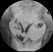
Image 5 Fluoroscopy (Frontal) ( update )
Caption
was changed:
Case 1 -: showing a bicornuate uterus
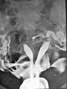
Image 6 Fluoroscopy ( update )
Caption
was changed:
Case 2 -: showing a septate uterus mimicking didelphys uterus
Image 7 Fluoroscopy ( update )

Caption
was changed:
Case 4 -: intra-uterine adhesions
Image 8 Fluoroscopy (HSG) ( update )

Caption
was changed:
Case 6 -: showing intra-uterine adhesions
Image 9 Fluoroscopy (Coronal) ( update )

Caption
was changed:
Case 3 -: showing a sub mucosal uterine leiomyoma
Image 10 Fluoroscopy (frontal) ( update )

Caption
was changed:
Case 5 -: showing hydrosalpinx


 Unable to process the form. Check for errors and try again.