
Lower limb radiography
Updates to Article Attributes
Body
was changed:
Lower limb radiography is the radiological investigation of the pelvis, hip joint, femur, knee joint, tibia, fibula, ankle joint, tarsal bones of the foot and metatarsals. It is often utilised in the context of trauma to rule out fractures and dislocations.
-<p><strong>Lower limb radiography</strong> is the radiological investigation of the <a title="Pelvis" href="/articles/pelvis-1">pelvis</a>, <a title="Hip joint" href="/articles/hip-joint-1">hip joint</a>, <a title="Femur" href="/articles/femur">femur</a>, <a title="Knee joint" href="/articles/knee-joint-1">knee joint</a>, tibia, fibula, <a title="Ankle joint" href="/articles/ankle-joint-2">ankle joint</a>, <a title="Tarsal bones" href="/articles/tarsal-bones">tarsal bones of the foot</a> and <a title="Metatarsals" href="/articles/metatarsals">metatarsals</a>. It is often utilised in the context of trauma to rule out fractures and dislocations. </p>- +<p><strong>Lower limb radiography</strong> is the radiological investigation of the <a href="/articles/pelvis-1">pelvis</a>, <a href="/articles/hip-joint-1">hip joint</a>, <a href="/articles/femur">femur</a>, <a href="/articles/knee-joint-1">knee joint</a>, tibia, fibula, <a href="/articles/ankle-joint-2">ankle joint</a>, <a href="/articles/tarsal-bones">tarsal bones of the foot</a> and <a href="/articles/metatarsals">metatarsals</a>. It is often utilised in the context of trauma to rule out fractures and dislocations. </p>
Images Changes:
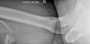
Image 6 X-ray (Horizontal beam lateral) ( update )
Position
was set to
.
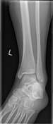
Image 7 X-ray (Frontal) ( update )

Position
was set to
.
Image 8 X-ray (Lateral) ( update )

Position
was set to
.
Image 9 X-ray (Horizontal beam lateral) ( update )

Position
was set to
.
Image 10 X-ray (Frontal) ( update )

Position
was set to
.
Image 11 X-ray (Lateral) ( update )

Position
was set to
.
Image 12 X-ray (Frontal) ( update )

Position
was set to
.
Image 13 X-ray (Mortice) ( update )
Position
was set to
.
Image 14 X-ray (Lateral) ( update )

Position
was set to
.
Image 15 X-ray (Frontal) ( update )

Position
was set to
.
Image 16 X-ray (Oblique) ( update )

Position
was set to
.
Image 17 X-ray (Lateral) ( update )

Position
was set to
.


 Unable to process the form. Check for errors and try again.