
MR enterography
Updates to Article Attributes
Body
was changed:
MR enterography is a new non-invasive technique for diagnosis of small bowel disorders.
Indications
The most common indication is to evaluate patients with Crohn's disease (CD).
Technique
Actual procedure will vary depending on institutional protocol/guidelines but below is a typical description 1, 2:
- patients should abstain from all food and drinks for 4-6 h prior to the study
- patients drink about 1-1.5 L of a 2.5% mannitol solution at regular intervals over a period of approximately 40 min prior to the study
- this solution acts as a hyperosmolar agent which draw fluid into the bowel
&and (biphasic) appears as low signal intensity on T1-weighted images and high signal intensity on T2-weighted images
- this solution acts as a hyperosmolar agent which draw fluid into the bowel
- scanning is ideally performed on a 1.5-T MRI scanner, using a phased array surface coil, either in the supine or prone position
MR protocol
- comprehensive MR examination of the small bowel usually requires axial and coronal both T1 and T2 weighted images
- high-resolution ultra-fast sequences such as true fast imaging with steady-state precession (true FISP) and HASTE sequences with and without fat suppression are usually used
- fat-suppressed three-dimensional (3D) T1-weighted breath-hold gradient-echo images of the abdomen and pelvis before and after intravenous gadolinium-based contrast material administration
Advantages
- no ionizing radiation
- high contrast resolution and multiplanar capability
- it can detect mural small bowel disorders with possible extramural complications and also allows the detection of the extra-intestinal solid organ pathologies
See also
-<p><strong>MR enterography</strong> is a new non-invasive technique for diagnosis of small bowel disorders.</p><h4>Indications</h4><p>The most common indication is to evaluate patients with <a href="/articles/crohns-disease-4">Crohn's disease</a> (CD).</p><h4>Technique</h4><p>Actual procedure will vary depending on institutional protocol/guidelines but below is a typical description <sup>1, 2</sup>:</p><ul>- +<p><strong>MR enterography</strong> is a new non-invasive technique for diagnosis of small bowel disorders.</p><h4>Indications</h4><p>The most common indication is to evaluate patients with <a href="/articles/crohn-disease-1">Crohn's disease</a> (CD).</p><h4>Technique</h4><p>Actual procedure will vary depending on institutional protocol/guidelines but below is a typical description <sup>1, 2</sup>:</p><ul>
-<li>patients drink about 1-1.5 L of a 2.5% mannitol solution at regular intervals over a period of approximately 40 min prior to the study<ul><li>this solution acts as a hyperosmolar agent which draw fluid into the bowel & (biphasic) appears as low signal intensity on T1-weighted images and high signal intensity on T2-weighted images</li></ul>- +<li>patients drink about 1-1.5 L of a 2.5% mannitol solution at regular intervals over a period of approximately 40 min prior to the study<ul><li>this solution acts as a hyperosmolar agent which draw fluid into the bowel and (biphasic) appears as low signal intensity on T1-weighted images and high signal intensity on T2-weighted images</li></ul>
Images Changes:
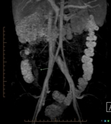
Image 2 MRI (MIP dynamic contrast) ( create )


 Unable to process the form. Check for errors and try again.