
Nasal bone
Updates to Article Attributes
Body
was changed:
The nasal bones are small paired oblong upper central facial bones placed side by side between the frontal processes of the maxilla, jointly forming the nasal ridge.
Gross anatomy
The nasal bone has two surfaces:
- external surface attaches to the procerus and nasalis muscles
- internal, which is transversely concave, with a groove for the anterior ethmoidal nerve
The four borders of the roughly quadrangular nasal bone are:
- superior: joins the frontal bone at the frontonasal suture
- inferior: free edge, continues as the lateral nasal cartilage
- lateral: articulates with the frontal process of the maxilla at the nasomaxillary suture
- medial: with a suture between it and its paired fellow at the internasal suture
Attachments
- musculotendinous
Ossification
The nasal bones ossify from a single centre which appears early in the third month within the cartilaginous nasal capsule, which can be assessed during the 12th-week prenatal scan for nasal bone appearance. Its absence is associated with Down syndrome.
-<li>inferior: free edge, continues as the <a title="Nasal cartilages" href="/articles/nasal-cartilages">lateral nasal cartilage</a>- +<li>inferior: free edge, continues as the <a href="/articles/nasal-cartilages">lateral nasal cartilage</a>
Images Changes:
Image 1 Diagram ( create )
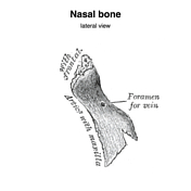
Image 2 Diagram ( create )
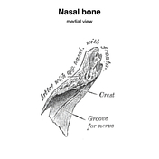
Image 3 Diagram ( create )

Image 4 X-ray (Lateral) ( update )

Position
was set to
.


 Unable to process the form. Check for errors and try again.