
Osteitis condensans ilii
Updates to Article Attributes
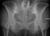
Osteitis condensans ilii is characterised by benign sclerosis of the ilium adjacent to the sacroiliac (SI) joint.
Pathology
The underlying etiology is believed to be mechanical stress across the SI joint. The fact that it is most often seen in women who have given birth supports this hypothesis, however also women who have not given birth and men can be affected 2-4.
Clinical presentation
It is usually asymptomatic but uncommonly may cause lower back pain, with a frequency about 1-2.5%4.
Pathology
The underlying etiology is believed to be mechanical stress across the SI joint. That it is most often seen in women who have given birth supports this hypothesis, however, women who have not given birth and men can be affected 2-4.
Radiographic features
Plain film and CT
Sclerosis of the ilial part of sacroiliac joint is seen, typically bilateral and triangular in shape 3-4. Lack of involvement of sacrum or joint space narrowing is considered diagnostic and thus may obviate need for further imaging 3. Unilateral disease has also been reported.
PrognosisTreatment and prognosis
It carries a benign prognosis and may even resolve spontaneously.
Differential diagnosis
The main differential diagnosis is a sacroiliitis, but with osteitis condensans ilii the SI joint is normal with no irregularity, erosions or loss of joint space.
-<p><strong>Osteitis condensans ilii</strong> is characterised by benign sclerosis of the ilium adjacent to the <a href="/articles/sacroiliac-joint">sacroiliac joint</a>. </p><h4>Pathology</h4><p>The underlying etiology is believed to be mechanical stress across the SI joint. The fact that it is most often seen in women who have given birth supports this hypothesis, however also women who have not given birth and men can be affected <sup>2-4</sup>. </p><h4>Clinical presentation</h4><p>It is usually asymptomatic but uncommonly may cause lower back pain, with a frequency about 1-2.5% <sup>4</sup>.</p><h4>Radiographic features</h4><h5>Plain film and CT</h5><p>Sclerosis of the ilial part of sacroiliac joint is seen, typically bilateral and triangular in shape <sup>3-4</sup>. Lack of involvement of sacrum or joint space narrowing is considered diagnostic and thus may obviate need for further imaging <sup>3</sup>. Unilateral disease has also been reported. </p><h4>Prognosis</h4><p>It carries a benign prognosis and may even resolve spontaneously. </p><h4>Differential diagnosis</h4><p>The main differential diagnosis is a <a href="/articles/differential-diagnosis-for-sacroiliac-joint-disease">sacroiliitis</a>, but with osteitis condensans ilii the SI joint is normal with no irregularity, erosions or loss of joint space.</p>- +<p><strong>Osteitis condensans ilii</strong> is characterised by benign sclerosis of the ilium adjacent to the <a href="/articles/sacroiliac-joint">sacroiliac (SI) joint</a>. </p><h4>Clinical presentation</h4><p>It is usually asymptomatic but uncommonly may cause lower back pain, with a frequency about 1-2.5% <sup>4</sup>.</p><h4>Pathology</h4><p>The underlying etiology is believed to be mechanical stress across the SI joint. That it is most often seen in women who have given birth supports this hypothesis, however, women who have not given birth and men can be affected <sup>2-4</sup>. </p><h4>Radiographic features</h4><h5>Plain film and CT</h5><p>Sclerosis of the ilial part of sacroiliac joint is seen, typically bilateral and triangular in shape <sup>3-4</sup>. Lack of involvement of sacrum or joint space narrowing is considered diagnostic and thus may obviate need for further imaging <sup>3</sup>. Unilateral disease has also been reported. </p><h4>Treatment and prognosis</h4><p>It carries a benign prognosis and may even resolve spontaneously. </p><h4>Differential diagnosis</h4><p>The main differential diagnosis is a <a href="/articles/sacroiliac-joint-disease-differential">sacroiliitis</a>, but with osteitis condensans ilii the SI joint is normal with no irregularity, erosions or loss of joint space.</p>
References changed:
- 2. Hutton CF. Osteitis condensans ilii. Br J Radiol. 1953;26 (309): 490-3. <a href="http://dx.doi.org/10.1259/0007-1285-26-309-490">doi:10.1259/0007-1285-26-309-490</a> - <a href="http://www.ncbi.nlm.nih.gov/pubmed/13081993">Pubmed citation</a><span class="auto"></span>
- 2.HUTTON CF. Osteitis condensans ilii. Br J Radiol. 1953;26 (309): 490-3. doi:10.1259/0007-1285-26-309-490 - Pubmed citation
Image 1 X-ray (Frontal) ( update )

Image 2 CT (bone window) ( update )

Image 3 X-ray ( update )

Image 4 MRI (T1) ( update )

Image 5 X-ray (Frontal) ( update )

Image 6 CT (bone window) ( update )

Image 7 X-ray ( update )

Image 8 X-ray (Frontal with annotations) ( update )


 Unable to process the form. Check for errors and try again.