
Osteoarthritis
Updates to Article Attributes
Osteoarthritis (OA), or degenerative joint disease (DJD), is the most common form of arthritis.
Terminology
Given osteoarthritis is not primarily an inflammatory process, as might be suggested by the suffix "itis", some authors prefer the term osteoarthrosis instead.
Pathology
Primary osteoarthritis is the less common variant and is characterised by the absence of an antecedent insult. There is a strong genetic component 5 with the disease primarily affecting middle-aged women.
Secondary osteoarthritis is more common, caused by abnormal mechanical forces (e.g. occupational stress, obesity) or by a previous joint insult (e.g. trauma, rheumatoid arthritis).
Radiographic features
Key radiographic features are joint space narrowing, sclerosis, and osteophytosis. If all three of these findings are not present, another diagnosis should be considered.
Joint space narrowing
- characteristically asymmetric
- least specific: present in many other pathological processes
Sclerosis
- sclerotic changes occur at joint margins
- frequently seen unless severe osteoporosis is present
Osteophytosis
- i.e. development of osteophytes
- common DJD finding
- will also be diminished in the setting of osteoporosis
- some osteophytes carry eponymous names, as discussed below
It affects the distal interphalangeal joints (Heberden nodes), the proximal interphalangeal joints (Bouchard nodes), (mnemonic H-D, B-P) and the base of the thumb in a bilaterally symmetric fashion. If it is not bilaterally symmetric, the diagnosis of primary osteoarthritis should be questioned.
Joint erosions
- several joints may exhibit degenerative erosions
- temporomandibular joint
- acromioclavicular joint
- sacroiliac joints
- symphysis pubis
Subchondral cyst
- also known as a geode
- cystic formations that occur around joints in a variety of disorders, including DJD, rheumatoid arthritis, calcium pyrophosphate dihydrate crystal deposition disease (CPPD) and avascular necrosis.
-<li>cystic formations that occur around joints in a variety of disorders, including DJD, <a href="/articles/rheumatoid-arthritis">rheumatoid arthritis</a>, <a href="/articles/calcium-pyrophosphate-dihydrate-deposition-disease">calcium pyrophosphate dihydrate crystal deposition disease (CPPD)</a> and <a href="/articles/avascular-necrosis">avascular necrosis</a>.</li>- +<li>cystic formations that occur around joints in a variety of disorders, including DJD, <a href="/articles/rheumatoid-arthritis">rheumatoid arthritis</a>, <a href="/articles/calcium-pyrophosphate-dihydrate-deposition-disease-1">calcium pyrophosphate dihydrate crystal deposition disease (CPPD)</a> and <a href="/articles/avascular-necrosis">avascular necrosis</a>.</li>
Image ( update )
Image 1 Diagram ( create )

Image 2 X-ray ( update )

Image 3 CT (bone window) ( update )
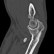
Image 5 MRI (PD fat sat) ( update )

Image 6 X-ray (Frontal) ( update )

Image 7 CT (bone window) ( update )

Image 8 X-ray (Frontal) ( update )

Image 11 X-ray (Frontal) ( update )

Image 12 X-ray (Frontal) ( update )


 Unable to process the form. Check for errors and try again.