
Peripheral pulmonary carcinoid tumor
Updates to Article Attributes
Peripheral pulmonary carcinoid tumour refer to a subtype of pulmonary carcinoid tumours that arise within the periphery of the lung. They are considered less common than the more centrally-located bronchial carcinoid tumours.
Clinical presentation
Many patients tend to be asymptomatic 2. Presentation with carcinoid syndrome is extremely rare 6.
Pathology
Peripheral pulmonary carcinoid tumours are considered a neuroendocrine tumour of the lung. They can be typical (well-differentiated - common) or atypical (more aggressive - uncommon).
Risk factors
- smoking: the rate of carcinoid tumours is similar between smokers and non-smokers, although there is an increased incidence of atypical subtype amongst smokers 5,6
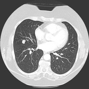
Radiographic features
HRCT/CT chestCT
Most are discovered as an incidental rounded solitary pulmonary nodule. The size at diagnosis can vary but is usually reported to be in the range of 10-30 mm 2. Many have a lobulated margin with an average Hounsfield value on postcontrast imaging of ~50 2. Imaging features are often non-specific and tissue diagnosis is essential in determining diagnosis.
Most peripheral carcinoid tumours tend to involve a subsegmental bronchus 2.
Nuclear medicine
FDG-PET
May have a sensitivity of around 75% 7. Although most cases will show uptake on an 18-FDG PET, up to a quarter of false negative has been described 2.
Galliun68-Octeotide-Octreotide-PET / 68Ga-DOTATATE
Usually avid and useful for diagnosis 8.
Differential diagnosis
See also
-<p><strong>Peripheral pulmonary carcinoid tumour</strong> refer to a subtype of <a href="/articles/pulmonary-carcinoid-tumours">pulmonary carcinoid tumours</a> that arise within the periphery of the lung. They are considered less common than the more centrally-located <a href="/articles/bronchial-carcinoid-tumour">bronchial carcinoid tumours</a>. </p><h4>Clinical presentation</h4><p>Many patients tend to be asymptomatic <sup> 2</sup>. Presentation with <a href="/articles/carcinoid-syndrome">carcinoid syndrome</a> is extremely rare <sup>6</sup>. </p><h4>Pathology</h4><p>Peripheral pulmonary carcinoid tumours are considered a <a href="/articles/neuroendocrine-tumour-of-the-lung">neuroendocrine tumour of the lung</a>. They can be typical (well-differentiated - common) or atypical (more aggressive - uncommon). </p><h5>Risk factors</h5><ul><li>smoking: rate of carcinoid tumours is similar between smokers and non-smokers, although there is increased incidence of atypical subtype amongst smokers <sup>5,6</sup>-</li></ul><h4>Radiographic features</h4><h5>HRCT/CT chest</h5><p>Most are discovered as an incidental rounded <a href="/articles/solitary-pulmonary-nodules">solitary pulmonary nodule</a>. The size at diagnosis can vary but is usually reported to be in the range of 10-30 mm <sup>2</sup>. Many have a lobulated margin with an average Hounsfield value on postcontrast imaging of ~50 <sup>2</sup>. Imaging features are often non-specific and tissue diagnosis is essential in determining diagnosis.</p><p>Most peripheral carcinoid tumours tend to involve a subsegmental bronchus <sup>2</sup>. </p><h5>Nuclear medicine</h5><h6>FDG-PET</h6><p>May have a sensitivity of around 75%<sup> 7</sup>.</p><h6>Galliun68-Octeotide-PET / <sup>68</sup>Ga-DOTATATE </h6><p>Usually avid and useful for diagnosis <sup>8</sup>.</p><h4>See also</h4><ul>-<li><a href="/articles/carcinoid-tumours-1">carcinoid tumours</a></li>-<li><a href="/articles/solitary-pulmonary-nodules">solitary pulmonary nodule</a></li>-<li><a href="/articles/pulmonary-tumourlet">pulmonary tumourlets</a></li>-</ul>- +<p><strong>Peripheral pulmonary carcinoid tumour</strong> refer to a subtype of <a href="/articles/pulmonary-carcinoid-tumours">pulmonary carcinoid tumours</a> that arise within the periphery of the lung. They are considered less common than the more centrally-located <a href="/articles/bronchial-carcinoid-tumour">bronchial carcinoid tumours</a>. </p><h4>Clinical presentation</h4><p>Many patients tend to be asymptomatic <sup> 2</sup>. Presentation with <a href="/articles/carcinoid-syndrome">carcinoid syndrome</a> is extremely rare <sup>6</sup>. </p><h4>Pathology</h4><p>Peripheral pulmonary carcinoid tumours are considered a <a href="/articles/neuroendocrine-tumour-of-the-lung">neuroendocrine tumour of the lung</a>. They can be typical (well-differentiated - common) or atypical (more aggressive - uncommon). </p><h5>Risk factors</h5><ul><li>smoking: the rate of carcinoid tumours is similar between smokers and non-smokers, although there is an increased incidence of atypical subtype amongst smokers <sup>5,6</sup>
- +</li></ul><h4>Radiographic features</h4><h5>CT</h5><p>Most are discovered as an incidental rounded <a href="/articles/solitary-pulmonary-nodules">solitary pulmonary nodule</a>. The size at diagnosis can vary but is usually reported to be in the range of 10-30 mm <sup>2</sup>. Many have a lobulated margin with an average Hounsfield value on postcontrast imaging of ~50 <sup>2</sup>. Imaging features are often non-specific and tissue diagnosis is essential in determining diagnosis.</p><p>Most peripheral carcinoid tumours tend to involve a subsegmental bronchus <sup>2</sup>. </p><h5>Nuclear medicine</h5><h6>FDG-PET</h6><p>May have a sensitivity of around 75%<sup> 7</sup>. Although most cases will show uptake on an 18-FDG PET, up to a quarter of false negative has been described <sup>2</sup>. </p><h6>Galliun68-Octreotide-PET / <sup>68</sup>Ga-DOTATATE </h6><p>Usually avid and useful for diagnosis <sup>8</sup>.</p><h4>Differential diagnosis</h4><p>See differentials for a <a title="Solitary pulmonary nodules" href="/articles/solitary-pulmonary-nodules">solitary</a><a title="Solitary pulmonary nodules" href="/articles/solitary-pulmonary-nodules"> pulmonary nodule</a>. </p>
Image 2 CT (lung window) ( create )


 Unable to process the form. Check for errors and try again.