
Pneumobilia
Updates to Article Attributes
Pneumobilia, also known as aerobilia, is accumulation of air in the biliary tree. It is important to distinguish pneumobilia from portal venous gas, the other type of branching hepatic gas. There are many causes of pneumobilia and clinical context is often important to distinguish between these 3.
Causes
- recent biliary instrumentation
- ERCP
- percutaneous or intraoperative cholangiography (small amount of air only)
- incompetent sphincter of Oddi
- sphincterotomy (~ 50% have pneumobilia at 1 year)
- following passage of a gallstone
- scarring eg. chronic pancreatitis
- drugs eg. atropine
- congenital
- biliary-enteric surgical anastomosis
- cholecytoenterostomy
- choledochoduodenostomy (with or without bile sump syndrome 2)
- Whipple procedure
-
spontaneous biliary-enteric fistula (cholecystoduodenal accounts for ~70% 3)
- gallstone ileus
- peptic ulcer disease
- traumatic
- neoplasm, eg. cholangiocarcinoma, ampullary cancer
- infection (rare)
- cholangitis
- emphysematous cholecystitis (usually gallbladder gas only, ~20% will have air in biliary tree also)
- liver abscess (if contains gas and communicates with biliary tree)
- bronchopleuralbiliary fistula (rare)
Radiographic features
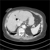
Pneumobilia is typically seen as linear branching air within the liver most prominent in central large calibre ducts as flow of bile pushes gas toward the hilum. This is in contrast to portal venous gas where peripheral small calibre branching air is usually seen due to flow of blood out from the hilum.
Plain film
Supine radiographs often demonstrate a sword-shaped lucency in the right paraspinal region representing gas from the common bile duct and the left hepatic duct. This has been termed the saber sign and is present in ~ 50% of patients with pneumobilia4.
Ultrasound
Ultrasound is very sensitive in detecting gas within the liver as it causes artefactartifact, specifically regions of high echogenicity with prominent shadowing or reverberation. The liver has been described as having a 'striped appearance'.
CT
Branching hepatic gas is easily appreciable on CT as branching air-density regions within the liver.
Differentiating between biliary and portal venous gas is usually achievable especially when intravenous contrast is used. Gas within the biliary tree tends to be more central, whereas gas within the portal venous system tends to be peripheral (pushed along by the blood). Also, biliary gas is ante-dependent, and typically fills the left lobe of the liver.
Differential diagnosis
The differential of pneumobilia is very limited :
-
portal venous gas
- patients usually ill (e.g. ischaemic bowel)
- gas more peripheral in liver
- Doppler imaging may help
- hepatic artery calcification (on ultrasound)
- often seen in those with chronic renal failure
- mimic pneumobilia on ultrasound 5
More importantly every attempt should be made at assessing the cause of pneumobilia, as a number of causative entities require prompt management (e.g. intestinal ischaemia).
See also
-</ul><h4>Radiographic features</h4><p>Pneumobilia is typically seen as linear branching air within the liver most prominent in central large calibre ducts as flow of bile pushes gas toward the hilum. This is in contrast to <a href="/articles/portal-venous-gas">portal venous gas</a> where peripheral small calibre branching air is usually seen due to flow of blood out from the hilum. </p><h5>Plain film</h5><p>Supine radiographs often demonstrate a sword-shaped lucency in the right paraspinal region representing gas from the common bile duct and the left hepatic duct. This has been termed the <a href="/articles/saber-sign-in-pneumobilia">saber sign</a> and is present in ~ 50% of patients with pneumobilia<sup>4</sup>.</p><h5>Ultrasound</h5><p>Ultrasound is very sensitive in detecting gas within the liver as it causes artefact, specifically regions of high echogenicity with prominent shadowing or reverberation. The liver has been described as having a 'striped appearance'.</p><h5>CT</h5><p>Branching hepatic gas is easily appreciable on CT as branching air-density regions within the liver.</p><p>Differentiating between biliary and portal venous gas is usually achievable especially when intravenous contrast is used. Gas within the biliary tree tends to be more central, whereas gas within the portal venous system tends to be peripheral (pushed along by the blood). Also, biliary gas is ante-dependent, and typically fills the left lobe of the liver.</p><h4>Differential diagnosis</h4><p>The differential of pneumobilia is very limited :</p><ul>- +</ul><h4>Radiographic features</h4><p>Pneumobilia is typically seen as linear branching air within the liver most prominent in central large calibre ducts as flow of bile pushes gas toward the hilum. This is in contrast to <a href="/articles/portal-venous-gas">portal venous gas</a> where peripheral small calibre branching air is usually seen due to flow of blood out from the hilum.</p><h5>Plain film</h5><p>Supine radiographs often demonstrate a sword-shaped lucency in the right paraspinal region representing gas from the common bile duct and the left hepatic duct. This has been termed the <a href="/articles/saber-sign-in-pneumobilia">saber sign</a> and is present in ~ 50% of patients with pneumobilia <sup>4</sup>.</p><h5>Ultrasound</h5><p>Ultrasound is very sensitive in detecting gas within the liver as it causes artifact, specifically regions of high echogenicity with prominent shadowing or reverberation. The liver has been described as having a 'striped appearance'.</p><h5>CT</h5><p>Branching hepatic gas is easily appreciable on CT as branching air-density regions within the liver.</p><p>Differentiating between biliary and portal venous gas is usually achievable especially when intravenous contrast is used. Gas within the biliary tree tends to be more central, whereas gas within the portal venous system tends to be peripheral (pushed along by the blood). Also, biliary gas is ante-dependent, and typically fills the left lobe of the liver.</p><h4>Differential diagnosis</h4><p>The differential of pneumobilia is very limited :</p><ul>
Image 1 Annotated image ( create )

Image 2 X-ray (Frontal) ( update )

Image 3 CT (Biliscopin) ( update )

Image 4 CT (non-contrast) ( update )

Image 5 CT (C+ portal venous phase) ( update )

Image 6 CT (renal cortical phase) ( update )


 Unable to process the form. Check for errors and try again.