
Pseudosubarachnoid hemorrhage
Updates to Article Attributes
Body
was changed:
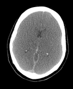
A pseudo-subarachnoid haemorrhage refers to apparent increased attenuation within the basal cisterns which simulates a true subarachnoid haemorrhage (SAH).
Pathology
Associations
- recent resuscitation from cardiopulmonary arrest:decreases in parenchyma attenuation due cerebral oedema and engorgement and dilatation of the superficial venous structures due an increased intracranial pressure
21,2 - severe meningitis: breakdown of the blood brain barrier allowing proteinaceous material to leak into the subarachnoid space3
- venous sinus thrombosis
- bilateral large subdural haemorrhage producing effacement of sulci and basilar cisterns and the false impression of blood in the subarachnoid space 5
Radiographic features
CT
- usually symmetrical density confined to basal cisterns (i.e. no sulcal density)
- 30-40 HU (compared with true acute SAH ~ 60HU)
- often seen with generalised cerebral oedema or basal cistern effacement
- the appearances thought due to combination of
- cisternal effacement
- distention +/- thrombosis of vessels
- adjacent brain hypoattenuation accentuating contrast difference
Given et al. reviewed 7 cases of generalised cerebral oedema accompanied by increased basal cisternal attenuation which were all found not to have subarachnoid blood at lumbar puncture or autopsy 1.
Differential diagnoses
- true subarachnoid haemorrhage
- acute leptomeningitis mimicking a subarachnoid haemorrhage 3
-<p>A <strong>pseudo-subarachnoid haemorrhage</strong> refers to apparent increased attenuation within the basal cisterns which simulates a true <a href="/articles/subarachnoid-haemorrhage">subarachnoid haemorrhage</a> (SAH).</p><h4>Pathology</h4><h5>Associations</h5><ul><li>recent resuscitation from cardiopulmonary arrest <sup>2</sup>-</li></ul><h4>Radiographic features</h4><h5>CT</h5><ul>- +<p>A <strong>pseudo-subarachnoid haemorrhage</strong> refers to apparent increased attenuation within the basal cisterns which simulates a true <a href="/articles/subarachnoid-haemorrhage">subarachnoid haemorrhage</a> (SAH).</p><h4>Pathology</h4><h5>Associations</h5><ul>
- +<li>recent resuscitation from cardiopulmonary arrest:<sup> </sup>decreases in parenchyma attenuation due <a href="/articles/cerebral-oedema-1">cerebral oedema</a> and engorgement and dilatation of the superficial venous structures due an <a href="/articles/increased-intracranial-pressure">increased intracranial pressure</a> <sup>1,2</sup>
- +</li>
- +<li>severe <a href="/articles/leptomeningitis">meningitis</a>: breakdown of the <a href="/articles/blood-brain-barrier">blood brain barrier</a> allowing proteinaceous material to leak into the <a href="/articles/subarachnoid-space">subarachnoid space</a> <sup>3</sup>
- +</li>
- +<li><a title="Dural venous sinus thrombosis" href="/articles/dural-venous-sinus-thrombosis">venous sinus thrombosis</a></li>
- +<li>bilateral large <a title="Subdural haemorrhage (SDH)" href="/articles/subdural-haemorrhage">subdural haemorrhage</a> producing effacement of sulci and basilar cisterns and the false impression of blood in the subarachnoid space <sup>5</sup>
- +</li>
- +</ul><h4>Radiographic features</h4><h5>CT</h5><ul>
-<li>often seen with generalised <a href="/articles/cerebral-oedema-1">cerebral oedema</a> or basal cistern effacement</li>- +<li>often seen with generalised cerebral oedema or basal cistern effacement</li>
References changed:
- 4. Opeskin K, Silberstein M. False positive diagnosis of subarachnoid haemorrhage on computed tomography scan. J Clin Neurosci. 2012;5 (4): 382-6. <a href="http://www.ncbi.nlm.nih.gov/pubmed/18639056">Pubmed citation</a><span class="auto"></span>
- 5. Rabinstein AA, Pittock SJ, Miller GM et-al. Pseudosubarachnoid haemorrhage in subdural haematoma. J. Neurol. Neurosurg. Psychiatr. 2003;74 (8): 1131-2. <a href="http://www.ncbi.nlm.nih.gov/pmc/articles/PMC1738620">Free text at pubmed</a> - <a href="http://www.ncbi.nlm.nih.gov/pubmed/12876252">Pubmed citation</a><span class="auto"></span>
Images Changes:
Image 1 CT (non-contrast) ( update )

Caption
was changed:
Case 1: oedema secondary to intracranial hypoxia
Image 2 CT (non-contrast) ( update )

Caption
was changed:
Case 2 : hyoxic or cerebral ischaemic event
Position
was set to
.
Image 3 CT (non-contrast) ( update )

Caption
was changed:
Case 3: hypoxic-ischaemic brain injury
Position
was set to
.
Image 4 CT (non-contrast) ( create )

Image 6 CT (non-contrast) ( update )
Position
was set to
.
Image 7 CT (non-contrast) ( update )

Caption
was changed:
Case 46
Position
was set to
.


 Unable to process the form. Check for errors and try again.