
Reticulonodular interstitial pattern
Updates to Article Attributes
Body
was changed:
A reticulonodular interstitial pattern is aan imaging descriptive term that can be used in thoracic radiographs or CT scans wherewhen are there is an overlap of reticular shadows with nodular shadows. This may be used to describe a regional pattern or a diffuse pattern throughout the lungs. It is a non specific imaging descriptor but can be seen in varied conditions such as
- silicosis
- pulmonary sarcoidosis 2
- berylliosis 2
- lymphangitis carcinomatosis 2
- hepatopulmonary syndrome - basal 4
- pneumocystis pneuomonia - can sometimes give a fine reticulonodular pattern 3
- bronchocentric granulomatosis 5
- pulmonary Langerhan cell histiocystosis 6
- lymphocytic interstitial pneumonitis 6
- Erdheim-Chester disease 1
-<p>A <strong>reticulonodular interstitial pattern</strong> is a descriptive term that can be used in thoracic radiographs or CT scans where are there is an overlap of <a title="reticular shadows" href="/articles/reticular-shadows">reticular shadows </a>with <a title="nodular shadows" href="/articles/nodular-shadows">nodular shadows</a>. This is a non specific imaging descriptor but can be seen in varied conditions such as </p><ul>- +<p>A <strong>reticulonodular interstitial pattern</strong> is an imaging descriptive term that can be used in thoracic radiographs or CT scans when are there is an overlap of <a href="/articles/reticular-shadows">reticular shadows </a>with <a href="/articles/nodular-shadows">nodular shadows</a>. This may be used to describe a regional pattern or a diffuse pattern throughout the lungs. It is a non specific imaging descriptor but can be seen in varied conditions such as</p><ul>
-<a title="Berylliosis involving lung" href="/articles/chronic-beryllium-lung-disease-1">berylliosis</a> <sup>2</sup>- +<a href="/articles/chronic-beryllium-lung-disease-1">berylliosis</a> <sup>2</sup>
-<a title="Erdheim-Chester disease" href="/articles/erdheim-chester-disease">Erdheim-Chester disease</a> <sup>1</sup>- +<a href="/articles/hepatopulmonary-syndrome">hepatopulmonary syndrome</a> - basal <sup>4</sup>
- +</li>
- +<li>
- +<a href="/articles/pneumocystis-pneumonia">pneumocystis pneuomonia</a> - can sometimes give a fine reticulonodular pattern <sup>3</sup>
- +</li>
- +<li>
- +<a href="/articles/bronchocentric-granulomatosis">bronchocentric granulomatosis</a> <sup>5</sup>
- +</li>
- +<li>
- +<a href="/articles/pulmonary-langerhans-cell-histiocytosis">pulmonary Langerhan cell histiocystosis</a> <sup>6</sup>
- +</li>
- +<li>
- +<a href="/articles/lymphocytic-interstitial-pneumonitis-1">lymphocytic interstitial pneumonitis</a> <sup>6</sup>
- +</li>
- +<li>
- +<a href="/articles/erdheim-chester-disease">Erdheim-Chester disease</a> <sup>1</sup>
References changed:
- 1. Chai G, Kaw G, Chuah K, Leong K, Chee C. Diffuse Reticulonodular Shadows: A Rare Manifestation of a Rare Disease. Chest. 2013;143(1):252-7. <a href="https://doi.org/10.1378/chest.11-2379">doi:10.1378/chest.11-2379</a> - <a href="https://www.ncbi.nlm.nih.gov/pubmed/23276850">Pubmed</a>
- 2. Oikonomou A & Prassopoulos P. Mimics in Chest Disease: Interstitial Opacities. Insights Imaging. 2013;4(1):9-27. <a href="https://doi.org/10.1007/s13244-012-0207-7">doi:10.1007/s13244-012-0207-7</a> - <a href="https://www.ncbi.nlm.nih.gov/pubmed/23247773">Pubmed</a>
- 3. Beigelman-Aubry C, Godet C, Caumes E. Lung Infections: The Radiologist's Perspective. Diagn Interv Imaging. 2012;93(6):431-40. <a href="https://doi.org/10.1016/j.diii.2012.04.021">doi:10.1016/j.diii.2012.04.021</a> - <a href="https://www.ncbi.nlm.nih.gov/pubmed/22658280">Pubmed</a>
- 4. McAdams H, Erasmus J, Crockett R, Mitchell J, Godwin J, McDermott V. The Hepatopulmonary Syndrome: Radiologic Findings in 10 Patients. AJR Am J Roentgenol. 1996;166(6):1379-85. <a href="https://doi.org/10.2214/ajr.166.6.8633451">doi:10.2214/ajr.166.6.8633451</a> - <a href="https://www.ncbi.nlm.nih.gov/pubmed/8633451">Pubmed</a>
- 5. Ohshimo S, Guzman J, Costabel U, Bonella F. Differential Diagnosis of Granulomatous Lung Disease: Clues and Pitfalls: Number 4 in the Series "Pathology for the Clinician" Edited by Peter Dorfmüller and Alberto Cavazza. Eur Respir Rev. 2017;26(145). <a href="https://doi.org/10.1183/16000617.0012-2017">doi:10.1183/16000617.0012-2017</a> - <a href="https://www.ncbi.nlm.nih.gov/pubmed/28794143">Pubmed</a>
- 6. Cosgrove G, Frankel S, Brown K. Challenges in Pulmonary Fibrosis. 3: Cystic Lung Disease. Thorax. 2007;62(9):820-29. <a href="https://doi.org/10.1136/thx.2004.031013">doi:10.1136/thx.2004.031013</a> - <a href="https://www.ncbi.nlm.nih.gov/pubmed/17726170">Pubmed</a>
Systems changed:
- Chest
Images Changes:
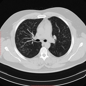
Image 1 CT (lung window) ( create )
Updates to Primarylink Attributes
Title
was added:
Reticulonodular interstitial pattern
Type
was set to
PrimaryLink.
Visible
was set to
.
Content
was set to
.
Updates to Synonym Attributes
Title
was added:
Reticulonodular pattern
Type
was set to
Synonym.
Visible
was set to
.
Content
was set to
.
Updates to Synonym Attributes
Title
was added:
Reticulonodular opacities
Type
was set to
Synonym.
Visible
was set to
.
Content
was set to
.


 Unable to process the form. Check for errors and try again.