
Solitary fibrous tumor
Updates to Article Attributes
A solitarySolitary fibrous tumour tumours (SFT) is aare a rare neoplasm of mesenchymal origin that comprises less thancomprise < 2% of allall soft tissue tumours tumours. The majority are benign The majority are benign, although up to 20% may be malignant.7
Epidemiology
Intrathoracic SFT usually presents in the 6th to 7th decades. Extrathoracic SFT occurs in men and women of all ages equally.7.
Clinical presentation
Although typically asymptomatic, it may present with symptoms and signs related to extrinsic compression of adjacent organs, or occasionally hypoglycemia4-7.
Pathology
Location
The majority of solitary fibrous tumours occur in an intrathoraciclocation but up to one third may occur in extrathoracic locations, and as such they may be encountered essentially anywhere, including 7:
- pleura (see solitary fibrous tumour of the pleura)
- spinal cord (see solitary fibrous tumour of the spinal cord) 2-3
- dura (see solitary fibrous tumour of the dura)
- head and neck
- extremities
- abdominal parenchymal organs
, - retroperitoneum
- peritoneum
- pelvic organs
Clinical presentation
Although typically asymptomatic, it may present with symptoms and signs related to extrinsic compression of adjacent organs, or occasionally hypoglycemia.4-7
Pathology
Histology
SFT most likely arises from submesothelial fibroblasts and is similar to a haemangiopericytoma.
At histology, a collagenous matrix with arrays of spindle cells is usually seen. Areas of necrosis, cystic or myxoid change, calcification, hemorrhage, increased vascularity, atypia, or malignanry may also be seen.7-8.
RaliographicRadiographic features
CT
CT reveals a wellwell-circumscribed, smoothsmooth, lobulated soft tissue mass that may contain scattered calcifications. Smaller tumors tend to enhance homogeneously, whereas larger lesions may have central tubular or rounded low-attenuation areas due to cystic or necrotic change.
MRI
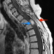
On MRI, benign solitary fibrous tumours usually has relatively homogeneous low to intermediate-to-intermediate signal intensity relativerelative to skeletal muscle on bothboth T1-weighted imaging and T2-weighted imaging because of fibrous tissue, as well as intense enhancement.
In addition, there may be areas of subacute haemorrhage that have high T1-weighted signal intensity, as well as nonenhancingas non-enhancing cystic or necrotic foci that are more heterogeneous and higher in T2-weighted signal intensity relative to thethe remainder of the tumour.
However, when a central focus of heterogeneity and variable contrast enhancement is identified in an SFT at CT or MRI, malignant degeneration should be considered.7.
Treatment and prognosis
-
abenign SFTless than 10cm< 10 cm in size typically has a favorable outcome with surgical resection alone. -
anSFTgreater than> 10 cm that also harbors malignant foci, and has positive surgical margins tends to have a poor prognosis, despite surgical treatment;- optimal adjuvant
therapytherapy forthe latterthis group is unknown, and close-interval follow-up is advised because there is an increased incidence of local recurrence.
- optimal adjuvant
-<p>A <strong>solitary fibrous tumour (SFT) </strong> is a rare neoplasm of mesenchymal origin that comprises less than 2% of all soft tissue tumours. The majority are benign, although up to 20% may be malignant.<sup>7</sup></p><h4>Epidemiology</h4><p>Intrathoracic SFT usually presents in the 6<sup>th</sup> to 7<sup>th</sup> decades. Extrathoracic SFT occurs in men and women of all ages equally.<sup>7</sup></p><h5>Location</h5><p>The majority of solitary fibrous tumours occur in an intrathoracic<strong> </strong>location but up to one third may occur in extrathoracic locations, and as such they may be encountered essentially anywhere, including <sup>7</sup>:</p><ul>- +<p><strong>Solitary fibrous tumours (SFT) </strong>are a rare neoplasm of mesenchymal origin that comprise < 2% of all soft tissue tumours. The majority are benign, although up to 20% may be malignant.</p><h4>Epidemiology</h4><p>Intrathoracic SFT usually presents in the 6<sup>th</sup> to 7<sup>th</sup> decades. Extrathoracic SFT occurs in men and women of all ages equally <sup>7</sup>.</p><h4>Clinical presentation</h4><p>Although typically asymptomatic, it may present with symptoms and signs related to extrinsic compression of adjacent organs, or occasionally <a href="/articles/hypoglycemia">hypoglycemia</a> <sup>4-7</sup>.</p><h4>Pathology</h4><h5>Location</h5><p>The majority of solitary fibrous tumours occur in an intrathoracic<strong> </strong>location but up to one third may occur in extrathoracic locations, and as such they may be encountered essentially anywhere, including <sup>7</sup>:</p><ul>
-<li>dura (see <a title="solitary fibrous tumour of the dura" href="/articles/solitary-fibrous-tumour-of-the-dura">solitary fibrous tumour of the dura</a>)</li>- +<li>dura (see <a href="/articles/solitary-fibrous-tumour-of-the-dura">solitary fibrous tumour of the dura</a>)</li>
-<li>abdominal parenchymal organs, </li>-<li>retroperitoneum</li>-<li>peritoneum </li>- +<li>abdominal parenchymal organs</li>
- +<li><a href="/articles/retroperitoneum">retroperitoneum</a></li>
- +<li><a href="/articles/peritoneum">peritoneum</a></li>
-</ul><h4>Clinical presentation</h4><p>Although typically asymptomatic, it may present with symptoms and signs related to extrinsic compression of adjacent organs, or occasionally <a href="/articles/hypoglycemia">hypoglycemia</a>.<sup>4-7</sup></p><h4>Pathology</h4><h5>Histology</h5><p>SFT most likely arises from submesothelial fibroblasts and is similar to a <a href="/articles/haemangiopericytoma-1">haemangiopericytoma</a>. </p><p>At histology, a collagenous matrix with arrays of spindle cells is usually seen. Areas of necrosis, cystic or myxoid change, calcification, hemorrhage, increased vascularity, atypia, or malignanry may also be seen.<sup>7-8</sup></p><h4>Raliographic features</h4><h5><strong>CT</strong></h5><p>CT reveals a well-circumscribed, smooth, lobulated soft tissue mass that may contain scattered calcifications. Smaller tumors tend to enhance homogeneously, whereas larger lesions may have central tubular or rounded low-attenuation areas due to cystic or necrotic change.</p><h5><strong>MRI</strong></h5><p>On MRI, benign solitary fibrous tumours usually has relatively homogeneous low to intermediate signal intensity relative to skeletal muscle on both T1-weighted imaging and T2-weighted imaging because of fibrous tissue, as well as intense enhancement. </p><p>In addition, there may be areas of subacute haemorrhage that have high T1-weighted signal intensity, as well as nonenhancing cystic or necrotic foci that are more heterogeneous and higher in T2-weighted signal intensity relative to the remainder of the tumour.</p><p>However, when a central focus of heterogeneity and variable contrast enhancement is identified in an SFT at CT or MRI, malignant degeneration should be considered.<sup>7</sup></p><h4>Treatment and prognosis</h4><ul>-<li>a benign SFT less than 10cm in size typically has a favorable outcome with surgical resection alone.</li>-<li>an SFT greater than 10 cm that also harbors malignant foci and has positive surgical margins tends to have a poor prognosis, despite surgical treatment; <br>optimal adjuvant therapy for the latter group is unknown, and close-interval follow-up is advised because there is an increased incidence of local recurrence.</li>- +</ul><h5>Histology</h5><p>SFT most likely arises from submesothelial fibroblasts and is similar to a <a href="/articles/haemangiopericytoma-1">haemangiopericytoma</a>. </p><p>At histology, a collagenous matrix with arrays of spindle cells is usually seen. Areas of necrosis, cystic or myxoid change, calcification, hemorrhage, increased vascularity, atypia, or malignanry may also be seen <sup>7-8</sup>.</p><h4>Radiographic features</h4><h5><strong>CT</strong></h5><p>CT reveals a well-circumscribed, smooth, lobulated soft tissue mass that may contain scattered calcifications. Smaller tumors tend to enhance homogeneously, whereas larger lesions may have central tubular or rounded low-attenuation areas due to cystic or necrotic change.</p><h5><strong>MRI</strong></h5><p>On MRI, benign solitary fibrous tumours usually has relatively homogeneous low-to-intermediate signal intensity relative to skeletal muscle on both T1-weighted imaging and T2-weighted imaging because of fibrous tissue, as well as intense enhancement. </p><p>In addition, there may be areas of subacute haemorrhage that have high T1-weighted signal intensity, as well as non-enhancing cystic or necrotic foci that are more heterogeneous and higher in T2-weighted signal intensity relative to the remainder of the tumour.</p><p>However, when a central focus of heterogeneity and variable contrast enhancement is identified in an SFT at CT or MRI, malignant degeneration should be considered <sup>7</sup>.</p><h4>Treatment and prognosis</h4><ul>
- +<li>benign SFT < 10 cm in size typically has a favorable outcome with surgical resection alone</li>
- +<li>SFT > 10 cm that also harbors malignant foci, and has positive surgical margins tends to have a poor prognosis, despite surgical treatment;<ul><li>optimal adjuvant therapy for this group is unknown, and close-interval follow-up is advised because there is an increased incidence of local recurrence</li></ul>
- +</li>
Image 1 CT (C+ CTPA) ( update )

Image 2 Annotated image ( update )


 Unable to process the form. Check for errors and try again.