
Spinal compression fracture
Updates to Article Attributes
Osteoporotic spinal compression fractures occur as a result of injury, commonly fall onto the buttock or pressure from normal activities, to the weakened vertebrae due to osteoporosis.
Epidemiology
They have a reported incidence of 1.2 per 1000 person-years after 85 years of age in the United States. However, they are largely unreported and are probably more common radiographically (present up to 14% of women older than 60 years in one study 1).
Clinical presentation
Vertebral fractures present with pain and loss of mobility.
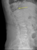
Radiographic features
Vertebral fractures require treatment when they are symptomatic, i.e. with pain and loss of mobility. This defines the role of the radiologist in making an accurate diagnosis.
The vertebral fracture should be diagnosed when there is a loss of height in the anterior, middle, or posterior dimension of the vertebral body that exceeds 20%. When in doubt, it is recommended that additional views or studies be advised for confirmation.
Osteoporotic spine fractures can be graded based on vertebral height loss as:
- mild: up to 20-25%
- moderate: 25-40%
- severe: >40%
Also see Genant classification of vertebral fractures
Acute vs chronic
Chronicity of the fracture indicates its temporal relationship with symptoms and hence is an important determination.
On conventional imaging, acute fracture signs include cortical breaking or impaction of trabeculae; in the absence of these signs fractures are chronic.
In uncertain cases, MRI signs of oedema (acute) and presence of radiotracer uptake on bone scintigraphy (acute) help decide the age of the fracture.
Treatment and prognosis
Management options include:
- non-surgical
- observation/bracing
- medications: bisphosphonates for osteoporosis
- surgical
-</ul><p>Also see <a title="Genant classification of vertebral fractures" href="/articles/genant-classification-of-vertebral-fractures">Genant classification of vertebral fractures</a></p><h5>Acute vs chronic</h5><p>Chronicity of the fracture indicates its temporal relationship with symptoms and hence is an important determination.</p><p>On conventional imaging, acute fracture signs include cortical breaking or impaction of trabeculae; in the absence of these signs fractures are chronic.</p><p>In uncertain cases, MRI signs of oedema (acute) and presence of radiotracer uptake on bone scintigraphy (acute) help decide the age of the fracture.</p><h4>Treatment and prognosis</h4><p>Management options include:</p><ul>- +</ul><p>Also see <a href="/articles/genant-classification-of-vertebral-fractures">Genant classification of vertebral fractures</a></p><h5>Acute vs chronic</h5><p>Chronicity of the fracture indicates its temporal relationship with symptoms and hence is an important determination.</p><p>On conventional imaging, acute fracture signs include cortical breaking or impaction of trabeculae; in the absence of these signs fractures are chronic.</p><p>In uncertain cases, MRI signs of oedema (acute) and presence of radiotracer uptake on bone scintigraphy (acute) help decide the age of the fracture.</p><h4>Treatment and prognosis</h4><p>Management options include:</p><ul>
Image 12 X-ray (Lateral) ( create )


 Unable to process the form. Check for errors and try again.