
Trochanteric fracture
Updates to Article Attributes
A trochanteric fracture is a fracture involving the greater and/or lesser trochanters of the femur.
Classification
Fractures in these regions can be classified as:
- intertrochanteric fracture
- pertrochanteric: intertrochanteric, involving both trochanters
- subtrochanteric fracture
- greater trochanteric avulsion fracture
- lesser trochanteric avulsion fracture
Inter-trochanteric fracture
They are subdivided according to the number of fragments, as follows:
- two-part linear intertrochanteric fracture stable
- three-part with comminution of lesser trochanter or greater trochanter
- four-part with comminution of both trochanters
- multi-part with comminution of both trochanters and intertrochanteric region
The Boyd and Griffin classification is based on the involvement of subtrochanteric region:
- type I: linear intertrochanteric
- type II: with comminution of trochanteric region
- type III: with comminution associated with the subtrochanteric component
- type IV: oblique fracture of the shaft with extension into the subtrochanteric region
The Tronzo classification is another classification system
- type I:incomplete fracture
- type II: no posteromedial comminution, lesser trochanter may be fractured
- type III: posteromedial comminution, the shaft is medially displaced with the neck beak impacted into it
- type III variant: type III combined with greater trochanter fracture
- type IV: posteromedial comminution, the shaft is laterally displaced
- type V: reverse obliquity
Subtrochanteric fracture
The Fielding classification of subtrochanteric fractures is based on the level of the subtrochanteric region through which the fracture extends:
- type I: at the level of the lesser trochanter (most common)
- type II: within the region 2.5 cm below the lesser trochanter
- type III: within the region 2.5 cm to 5 cm below the lesser trochanter (least common)
The Zickel classification (modified from Fielding) of subtrochanteric fractures takes into consideration the level and obliquity of the fracture line as well as the presence or absence of comminution.
-
type I: short oblique
- linear
- comminuted
-
type II: long oblique
- linear
- comminuted
-
type III: transverse
- high level
- low level
Treatment and prognosis
Sub-trochanteric fractures generally have a good prognosis due to the good supply of blood and adequate collateral circulation to this region of the femur with a low incidence of avascular necrosis and non-union. Post-operative infection, however, is a potentially serious complication.
-<p>A<strong> trochanteric fracture</strong> is a fracture involving the greater and/or lesser trochanters of the <a href="/articles/femur">femur</a>.</p><h4>Classification</h4><p>Fractures in these regions can be classified as:</p><ul>-<li><a href="/articles/intertrochanteric-fracture">intertrochanteric fracture</a></li>-<li>-<a href="/articles/pertrochanteric-fracture">pertrochanteric</a>: intertrochanteric, involving both trochanters</li>-<li><a href="/articles/subtrochanteric-fracture">subtrochanteric fracture</a></li>-<li><a href="/articles/greater-trochanteric-avulsion-fracture">greater trochanteric avulsion fracture</a></li>-<li><a href="/articles/lesser-trochanteric-avulsion-fracture">lesser trochanteric avulsion fracture</a></li>-</ul><h5>Inter-trochanteric fracture</h5><p>They are subdivided according to the number of fragments, as follows:</p><ul>-<li>two-part linear intertrochanteric fracture stable</li>-<li>three-part with comminution of lesser trochanter or greater trochanter</li>-<li>four-part with comminution of both trochanters</li>-<li>multi-part with comminution of both trochanters and intertrochanteric region</li>-</ul><p>The <strong>Boyd and Griffin classification</strong> is based on the involvement of subtrochanteric region:</p><ul>-<li>-<strong>type I</strong>: linear intertrochanteric</li>-<li>-<strong>type II</strong>: with comminution of trochanteric region</li>-<li>-<strong>type III</strong>: with comminution associated with the subtrochanteric component</li>-<li>-<strong>type IV</strong>: oblique fracture of the shaft with extension into the subtrochanteric region</li>-</ul><p>The <strong>Tronzo classification </strong>is another classification system</p><ul>-<li>-<strong>type I</strong>:<strong> </strong>incomplete fracture</li>-<li>-<strong>type II</strong>: no posteromedial comminution, lesser trochanter may be fractured</li>-<li>-<strong>type III</strong>: posteromedial comminution, the shaft is medially displaced with the neck beak impacted into it</li>-<li>-<strong>type III variant</strong>: type III combined with greater trochanter fracture</li>-<li>-<strong>type IV</strong>: posteromedial comminution, the shaft is laterally displaced</li>-<li>-<strong>type V</strong>: reverse obliquity</li>-</ul><h5>Subtrochanteric fracture</h5><p>The <strong>Fielding</strong> <strong>classification</strong> of subtrochanteric fractures is based on the level of the subtrochanteric region through which the fracture extends:</p><ul>-<li>-<strong>type I</strong>: at the level of the lesser trochanter (most common)</li>-<li>-<strong>type II</strong>: within the region 2.5 cm below the lesser trochanter</li>-<li>-<strong>type III</strong>: within the region 2.5 cm to 5 cm below the lesser trochanter (least common)</li>-</ul><p>The <strong>Zickel</strong> <strong>classification</strong> (modified from Fielding) of subtrochanteric fractures takes into consideration the level and obliquity of the fracture line as well as the presence or absence of comminution.</p><ul>-<li>-<strong>type I</strong>: short oblique<ul>-<li>linear</li>-<li>comminuted</li>-</ul>-</li>-<li>-<strong>type II</strong>: long oblique<ul>-<li>linear</li>-<li>comminuted</li>-</ul>-</li>-<li>-<strong>type III</strong>: transverse<ul>-<li>high level</li>-<li>low level</li>-</ul>-</li>- +<p>A<strong> trochanteric fracture</strong> is a fracture involving the greater and/or lesser trochanters of the <a href="/articles/femur">femur</a>.</p><h4>Classification</h4><p>Fractures in these regions can be classified as:</p><ul>
- +<li><a href="/articles/intertrochanteric-fracture">intertrochanteric fracture</a></li>
- +<li>
- +<a href="/articles/pertrochanteric-fracture">pertrochanteric</a>: intertrochanteric, involving both trochanters</li>
- +<li><a href="/articles/subtrochanteric-fracture">subtrochanteric fracture</a></li>
- +<li><a href="/articles/greater-trochanteric-avulsion-fracture">greater trochanteric avulsion fracture</a></li>
- +<li><a href="/articles/lesser-trochanteric-avulsion-fracture">lesser trochanteric avulsion fracture</a></li>
- +</ul><h5>Inter-trochanteric fracture</h5><p>They are subdivided according to the number of fragments, as follows:</p><ul>
- +<li>two-part linear intertrochanteric fracture stable</li>
- +<li>three-part with comminution of lesser trochanter or greater trochanter</li>
- +<li>four-part with comminution of both trochanters</li>
- +<li>multi-part with comminution of both trochanters and intertrochanteric region</li>
- +</ul><p>The <strong>Boyd and Griffin classification</strong> is based on the involvement of subtrochanteric region:</p><ul>
- +<li>
- +<strong>type I</strong>: linear intertrochanteric</li>
- +<li>
- +<strong>type II</strong>: with comminution of trochanteric region</li>
- +<li>
- +<strong>type III</strong>: with comminution associated with the subtrochanteric component</li>
- +<li>
- +<strong>type IV</strong>: oblique fracture of the shaft with extension into the subtrochanteric region</li>
- +</ul><p>The <strong>Tronzo classification </strong>is another classification system</p><ul>
- +<li>
- +<strong>type I</strong>:<strong> </strong>incomplete fracture</li>
- +<li>
- +<strong>type II</strong>: no posteromedial comminution, lesser trochanter may be fractured</li>
- +<li>
- +<strong>type III</strong>: posteromedial comminution, the shaft is medially displaced with the neck beak impacted into it</li>
- +<li>
- +<strong>type III variant</strong>: type III combined with greater trochanter fracture</li>
- +<li>
- +<strong>type IV</strong>: posteromedial comminution, the shaft is laterally displaced</li>
- +<li>
- +<strong>type V</strong>: reverse obliquity</li>
- +</ul><h5>Subtrochanteric fracture</h5><p>The <strong>Fielding</strong> <strong>classification</strong> of subtrochanteric fractures is based on the level of the subtrochanteric region through which the fracture extends:</p><ul>
- +<li>
- +<strong>type I</strong>: at the level of the lesser trochanter (most common)</li>
- +<li>
- +<strong>type II</strong>: within the region 2.5 cm below the lesser trochanter</li>
- +<li>
- +<strong>type III</strong>: within the region 2.5 cm to 5 cm below the lesser trochanter (least common)</li>
- +</ul><p>The <strong>Zickel</strong> <strong>classification</strong> (modified from Fielding) of subtrochanteric fractures takes into consideration the level and obliquity of the fracture line as well as the presence or absence of comminution.</p><ul>
- +<li>
- +<strong>type I</strong>: short oblique<ul>
- +<li>linear</li>
- +<li>comminuted</li>
- +</ul>
- +</li>
- +<li>
- +<strong>type II</strong>: long oblique<ul>
- +<li>linear</li>
- +<li>comminuted</li>
- +</ul>
- +</li>
- +<li>
- +<strong>type III</strong>: transverse<ul>
- +<li>high level</li>
- +<li>low level</li>
- +</ul>
- +</li>
Image ( destroy )
Image ( destroy )
Image 1 Diagram ( update )

Image 2 Diagram (Tronzo classification (blue)) ( create )

Image 3 X-ray (Frontal) ( update )
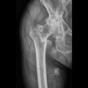
Image 4 X-ray ( update )

Image 5 X-ray (Frontal) ( update )

Image 6 X-ray (Frontal) ( update )

Image 7 X-ray (Oblique) ( update )

Image 8 X-ray (Frontal) ( update )

Image 9 X-ray (Frontal) ( update )

Image 10 MRI (T2 fat sat) ( update )


 Unable to process the form. Check for errors and try again.