
Adenoid cystic tumor of palate
Diagnosis certain
Updates to Study Attributes
Findings
was changed:
OPG
OPG. There is no bone abnormality seen within the mandible. There is an area of extensive bone destruction involving the maxilla on its left lateral side. These appearances are suspicious for bone destruction secondary to a probable squamous cell carcinoma.
Updates to Study Attributes
Findings
was changed:
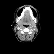
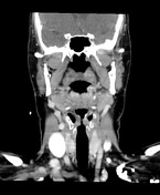
CT Neck
Soft tissue mass destroying the maxillary alveolus and hard palate.
There is involement of the left maxillary antrum and extentsion posteriorly into the pterygopalatine fossa.
No No enlarged cervical nodes are seen
Images Changes:
Image CT (C+ delayed) ( update )
Specifics
changed from C+ portal venous phase to C+ delayed.
Image CT (C+ delayed) ( update )
Specifics
changed from C+ portal venous phase to C+ delayed.
Updates to Case Attributes
Body
was changed:
Adenoid cystic tumour of the palate on histology.
-<p>Adenoid cystic tumour of the palate on histology.</p><p> </p>- +<p>Adenoid cystic tumour of the palate on histology.</p>
Systems changed:
- Oncology


 Unable to process the form. Check for errors and try again.