
Branchial cleft cyst
Diagnosis almost certain
Updates to Case Attributes
Age
changed from 45 to 45 years.
Body
was added:
Imaging findings of a neck cystic lesion with the classic location for a second branchial cleft cyst. No signs of infection or complications identified.
- +<p>Imaging findings of a neck cystic lesion with the classic location for a <a title="Second branchial cleft cyst" href="/articles/second-branchial-cleft-cyst">second branchial cleft cyst</a>. No signs of infection or complications identified. </p>
Updates to Study Attributes
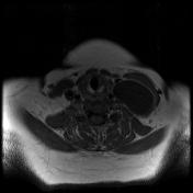
Findings
was added:
A well-defined cystic lesion in the left side of the neck deep to the left sternomastoid muscle with clear fluid content and a thin enhancing wall. It is lateral to the carotid sheath and posterior to the left submandibular gland.
Images Changes:
Image MRI (T2 fat sat) ( update )

Stack
was set to
.
Single Or Stack Root
was set to
.
Image MRI (T2 fat sat) ( update )

Stack
was set to
.
Single Or Stack Root
was set to
.
Perspective
was set to
Axial.
Image MRI (T2) ( update )

Perspective
was set to
Coronal.
Image MRI (T1) ( update )

Perspective
was set to
Axial.
Image MRI (T1 C+) ( update )
Perspective
was set to
Axial.


 Unable to process the form. Check for errors and try again.