
Bronchioloalveolar carcinoma
Updates to Case Attributes
Chest x-ray demonstrates extensive and widespread fluffy airspace opacities, with almost complete white-out of the mid and lower zones on the left.
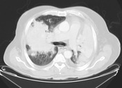
CT confirms extensive airspace opacities with numerous air-bronchograms. No pleural effusions or significantevidence of mediastinal adenopathy.
Sputum, right and left main bronchus lavage were positive for malignant cells consistent ofwith carcinoma, thought true cut. A Tru-cut biopsy was suggested by the pathologist to confirm the diagnosis of bronchoalveolar carcinoma, the patient's condition didmeant that this was not permit for thispossible.
-<p>Chest x-ray demonstrates extensive and widespread fluffy airspace opacities, with almost complete white-out of the mid and lower zones on the left.</p><p>CT confirms extensive airspace opacities with numerous air-bronchograms. No pleural effusions or significant adenopathy.</p><p>Sputum, right and left main bronchus lavage were positive for malignant cells consistent of carcinoma, thought true cut biopsy was suggested by the pathologist to confirm the diagnosis of <a href="/articles/adenocarcinoma-in-situ-minimally-invasive-adenocarcinoma-and-invasive-adenocarcinoma-of-lung">bronchoalveolar carcinoma</a>, the patient condition did not permit for this.</p>- +<p>Chest x-ray demonstrates extensive and widespread fluffy airspace opacities, with almost complete white-out of the mid and lower zones on the left.</p><p>CT confirms extensive airspace opacities with numerous air-bronchograms. No pleural effusions or evidence of mediastinal adenopathy.</p><p>Sputum, right and left main bronchus lavage were positive for malignant cells consistent with carcinoma. A Tru-cut biopsy was suggested by the pathologist to confirm the diagnosis of <a href="/articles/adenocarcinoma-in-situ-minimally-invasive-adenocarcinoma-and-invasive-adenocarcinoma-of-lung">bronchoalveolar carcinoma</a>, the patient's condition meant that this was not possible.</p>
Updates to Study Attributes
Widespread fluffy airspace opacities, with almost complete white-out of the mid and lower zones on the left.
Image X-ray (Frontal) ( update )

Updates to Study Attributes
Extensive airspace opacities with numerous air bronchograms. Trace of pleural fluid on the left only. No significant nodal enlargement.
Image CT (C+ arterial phase) ( update )

Image CT (lung window) ( update )


 Unable to process the form. Check for errors and try again.