
Cavernous malformation with DVA
Diagnosis certain
Updates to Case Attributes
Title
was changed:
Cavernous malformation with DVA
Age
was set to
Young adult.
Body
was changed:
A young female patient brain MRI demonstratesTypical appearances of a cavernous malformation. Susceptibility sequence failed to detect other similar lesions (Zabramski type 2) with an associated DVA, a very common finding.
-<p>A young female patient brain MRI demonstrates a right pons popcorn lesion with surrounding hemosidren ring with complex signal intensity due to blood product of varying age. The lesion is consistent with <a href="/articles/cerebral-cavernous-malformation" title="Cerebral cavernous malformation">cavernous malformation</a>. Susceptibility sequence failed to detect other similar lesions.</p>- +<p>Typical appearances of a <a title="Cerebral cavernous malformation" href="/articles/cerebral-cavernous-venous-malformation">cavernous malformation</a> (<a title="Zabramski classification of cerebral cavernous malformations" href="/articles/zabramski-classification-of-cerebral-cavernous-malformations">Zabramski</a> type 2) with an associated <a title="Developmental venous anomaly (DVA)" href="/articles/developmental-venous-anomaly">DVA</a>, a very common finding.</p>
Diagnostic Certainty
was set to
.
Updates to Study Attributes
Findings
was added:
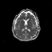
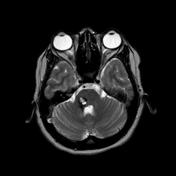
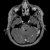
MRI demonstrates a right pontine popcorn lesion with a complete surrounding hemosiderin ring with complex signal intensity due to blood product of varying age. The lesion is consistent with a cavernous malformation. Note the presence of an associated developmental venous anomaly seen only on T1 C+ sequence, draining the right cerebellar hemisphere. Susceptibility sequence failed to detect other similar lesions.
Images Changes:
Image MRI (ADC) ( update )
Perspective
was set to
Axial.
Image MRI (FLAIR) ( update )

Perspective
was set to
Axial.
Description
was removed:
Image MRI (DWI) ( update )

Stack
was set to
.
Single Or Stack Root
was set to
.
Image MRI (DWI) ( update )

Stack
was set to
.
Single Or Stack Root
was set to
.
Perspective
was set to
Axial.
Specifics
was set to
DWI.
Description
was removed:
Image MRI (Gradient Echo) ( update )

Stack
was set to
.
Single Or Stack Root
was set to
.
Perspective
was set to
Axial.
Specifics
was set to
Gradient Echo.
Description
was removed:
Image MRI (Gradient Echo) ( update )

Stack
was set to
.
Single Or Stack Root
was set to
.
Image MRI (T2) ( update )
Stack
was set to
.
Single Or Stack Root
was set to
.
Perspective
was set to
Axial.
Description
was removed:
Image MRI (T2) ( update )

Stack
was set to
.
Single Or Stack Root
was set to
.
Image MRI (T1) ( update )

Perspective
was set to
Axial.
Description
was removed:
Image MRI (T2) ( update )

Perspective
was set to
Coronal.
Stack
was set to
.
Single Or Stack Root
was set to
.
Image MRI (T2) ( update )

Stack
was set to
.
Single Or Stack Root
was set to
.
Description
was removed:
Image MRI (T1 C+ fat sat) ( update )
Perspective
was set to
Axial.
Specifics
was set to
T1 C+ fat sat.
Description
was removed:


 Unable to process the form. Check for errors and try again.