
Cerebral toxoplasmosis
Updates to Study Attributes
A 24 x 22 x 23 mm mass is demonstrated centred in the right basal nuclei with an irregularly thick contrast enhancing rim. Significant surrounding T2/FLAIR high signal extends via the cerebral peduncle into the midbrain and inferiorly to the right cerebral peduncle which is expanded. There is moderate regional mass effect with effacement of the frontotemporal sulci and effacement of the right lateral ventricle, resulting bowing of the septum pellucidum after 4 mm to the left.
The central portion of the mass is mildly hypointense to grey matter on T1-weighted imaging and hyperintense on T2-weighted imaging with poor suppression on FLAIR. Diffusion weighted imaging demonstrates patchy high signal predominantly at the peripheral portion of the mass and centrally decreased ADC. There is mildly increased rCBV in the rim of the mass. Spectroscopy demonstrates metabolite depletion and prominent lactate peak.
Conclusion:
RimIn an immunocompetent patient, this rim-enhancing mass centered in the right basal nuclei has features favouring a high-grade glioma. Features are not typical of a pyogenic abscess however abscesses secondary to atypical organisms may(e.g. Toxoplasmosis) may have this appearance; HIV / immunosuppressed status needs to be assessed. A less likely diagnosis is lymphoma, which may have this appearance in an immunocompromised patient.
Image MRI (Echo planar) ( update )

Image MRI (T1 C+) ( update )

Image MRI (FLAIR) ( update )

Image MRI (T1) ( update )
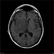
Image MRI (DWI) ( update )
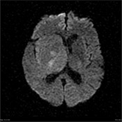
Image MRI (ADC) ( update )
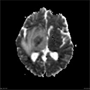
Image MRI (T1) ( update )

Image MRI (T1) ( update )

Image MRI (T1 C+) ( update )
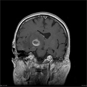
Image 1 MRI (T2) ( update )

Image 2 MRI (FLAIR) ( create )

Image 3 MRI (T1) ( create )
Image 4 MRI (T1 C+) ( create )
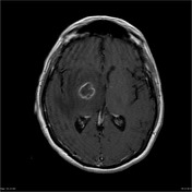
Image 5 MRI (T1) ( create )
Image 6 MRI (T1 C+) ( create )
Image 7 MRI (DWI) ( create )
Image 8 MRI (ADC) ( create )
Image 9 MRI (MRS) ( update )

Image 10 MRI (CBV) ( update )
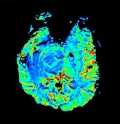
Image 11 MRI (Echo planar) ( create )


 Unable to process the form. Check for errors and try again.