
Chondroblastoma
Diagnosis certain
Updates to Study Attributes
Findings
was changed:
Ill-defined lucency within the proximal humeral epiphysis.
Images Changes:
Image X-ray (Frontal) ( update )

Description
was removed:
Single Or Stack Root
was set to
.
Perspective
was set to
Frontal.
Updates to Study Attributes
Findings
was changed:
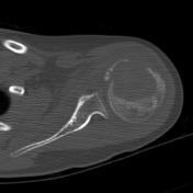
CT confirms an aggressive lytic lesion of the proximal humeral epiphysis with cortical destruction and ill-defined surrounding sclerosis.
Images Changes:
Image CT (bone window) ( update )
Perspective
was set to
Axial.
Single Or Stack Root
was set to
.
Specifics
was set to
bone window.
Description
was removed:
Image CT (bone window) ( update )

Perspective
was set to
Coronal.
Single Or Stack Root
was set to
.
Specifics
was set to
bone window.
Description
was removed:
Updates to Study Attributes
Findings
was changed:
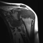
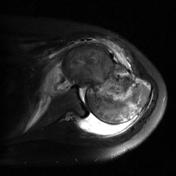
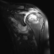
MRI demonstrates an aggressive lesion that is T1 hypointense and T2 hyperintense.
Images Changes:
Image MRI (STIR) ( update )

Perspective
was set to
Coronal.
Single Or Stack Root
was set to
.
Description
was removed:
Image MRI (T1) ( update )
Perspective
was set to
Coronal.
Single Or Stack Root
was set to
.
Description
was removed:
Image MRI (T2 fat sat) ( update )
Perspective
was set to
Axial.
Single Or Stack Root
was set to
.
Description
was removed:
Image MRI (T2 fat sat) ( update )
Perspective
was set to
Coronal.
Single Or Stack Root
was set to
.
Description
was removed:
Image 2 MRI (STIR) ( update )
Position
was set to
.
Updates to Case Attributes
Diagnostic Certainty
was set to
.
Systems changed:
- Oncology


 Unable to process the form. Check for errors and try again.