
Chondrosarcoma - sphenoid wing
Diagnosis almost certain
Updates to Case Attributes
Title
was changed:
Chondrosarcoma - sphenoid wing - large
Age
changed from 57 to 55 years.
Body
was changed:
histologyThe patient went on to have a resection. Histology confirmed chondrosarcoma in a 57 year old malechondrosarcoma.
-<p>histology confirmed chondrosarcoma in a 57 year old male. </p>- +<p>The patient went on to have a resection. Histology confirmed a <a title="Chondrosarcoma" href="/articles/chondrosarcoma">chondrosarcoma</a>.</p>
Presentation
was added:
Headaches.
Updates to Link Attributes
Title
was removed:
Type
was removed.
Visible
was set to
.
Updates to Primarylink Attributes
Updates to Study Attributes
Findings
was changed:
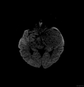
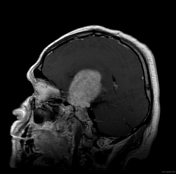
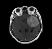
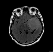
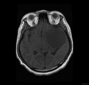
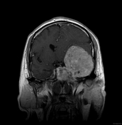
LeftSelected MRI images demonstrate a large left sphenoid wing extra axial-axial mass that has very high T2 signal and vivid contrast enhancement. The adjacent brain is displaced with minimal oedema.
The off-midline location and very high T2 signal suggest the diagnosis of chondrosarcoma.
Images Changes:
Image MRI (DWI) ( update )
Description
was removed:
Image MRI (T2) ( update )

Description
was removed:
Image MRI (T1 C+) ( update )
Description
was removed:
Image MRI (T1 C+) ( update )
Description
was removed:
Image MRI (FLAIR) ( update )
Description
was removed:
Image MRI (T1) ( update )
Description
was removed:
Image MRI (T1 C+) ( update )
Description
was removed:
Image 1 MRI (T2) ( update )
Position
was set to
.
Image 2 MRI (FLAIR) ( update )
Position
was set to
.
Image 3 MRI (T1) ( update )
Position
was set to
.
Image 4 MRI (T1 C+) ( update )
Position
was set to
.
Image 5 MRI (T1 C+) ( update )
Position
was set to
.
Image 6 MRI (T1 C+) ( update )
Position
was set to
.


 Unable to process the form. Check for errors and try again.